. 2003 Dec;163(6):2233-45.
doi: 10.1016/S0002-9440(10)63581-X.

B lymphocyte-specific c-Myc expression stimulates early and functional expansion of the vasculature and lymphatics during lymphomagenesis

Affiliations

B lymphocyte-specific c-Myc expression stimulates early and functional expansion of the vasculature and lymphatics during lymphomagenesis

Alanna Ruddell et al. Am J Pathol. 2003 Dec.

Abstract

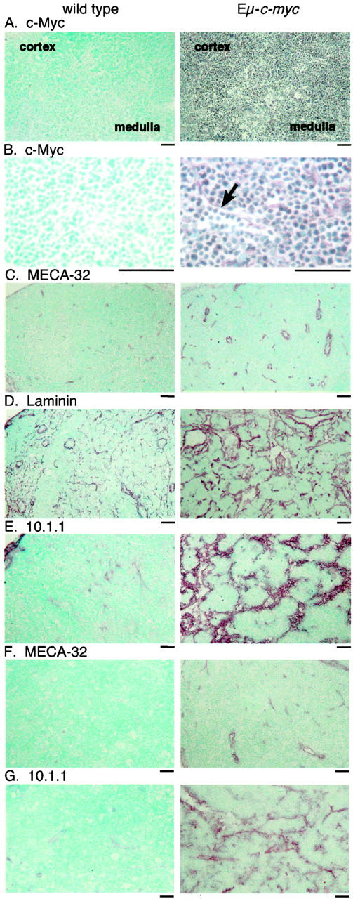
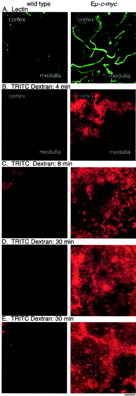
Expression of the c-myc proto-oncogene is deregulated in many human cancers. We examined the role of c-Myc in stimulating angiogenesis and lymphangiogenesis in a highly metastatic murine model of Burkitt's lymphoma (E micro -c-myc), where c-Myc is expressed exclusively in B lymphocytes. Immunohistochemical analysis of bone marrow and lymph nodes from young (preneoplastic) E micro -c-myc transgenic mice revealed increased growth of blood vessels, which are functional by dye flow assay. Lymphatic sinuses also increased in size and number within the lymph nodes, as demonstrated by immunostaining for with a lymphatic endothelial marker 10.1.1. The 10.1.1 antibody recognizes VEGFR-2- and VEGFR-3-positive lymphatic sinuses and vessels within lymph nodes, and also recognizes lymphatic vessels in other tissues. Subcutaneously injected dye traveled more efficiently through draining lymph nodes in E micro -c-myc mice, indicating that these hypertrophic lymphatic sinuses increase lymph flow. Purified B lymphocytes and lymphoid tissues from E micro -c-myc mice expressed increased levels of vascular endothelial growth factor (VEGF) by immunohistochemical or immunoblot assays, which could promote blood and lymphatic vessel growth through interaction with VEGFR-2, which is expressed on the endothelium of both vessel types. These results indicate that constitutive c-Myc expression stimulates angiogenesis and lymphangiogenesis, which may promote the rapid growth and metastasis of c-Myc-expressing cancer cells, respectively.


Figures

Figure 1.
Figure 1.
c-Myc-expressing B cells increase the functional vasculature in bone marrow of Eμ-c-myc mice. A: Bone marrow cryosections immunostained for c-Myc show accumulation of c-Myc-expressing B-cell progenitors within Eμ-c-myc marrow from 4-week-old mice, while c-Myc-expressing cells are rare in wild-type littermate marrow. B: Paraffin sections stained for MECA-32 show increased number and size of small vessels (arrows) in 8-week-old Eμ-c-myc mice, relative to littermate controls. C: Paraffin sections stained for laminin reveal increased venous sinuses (arrows) and small blood vessels in Eμ-c-myc mice. D: Cryosections from FITC-lectin-injected mice were immunostained with Texas Red-labeled laminin antibody. The laminin-coated venous sinuses and blood vessels are functional, as the FITC- and Texas Red-laminin immunostaining decorate the same vessels. E: 50-μm bone marrow cryosections from FITC-lectin-injected mice demonstrate increased functional FITC-labeled vessels in Eμ-c-myc mice relative to littermates. F: As a control, 50-μm kidney cryosections from lectin-injected mice show similar blood flow in glomeruli of control and Eμ-c-myc mice. Panels are shown at ×200 magnification; scale bars are 50 μm.
Figure 2.
Figure 2.
Functional blood vessel density is increased in bone marrow from Eμ-c-myc mice. The number of discrete FITC-lectin-labeled vessels were counted per 400-μm field, in 50-μm bone marrow cryosections from five pairs of wild-type and Eμ-c-myc FITC lectin-injected mice. Mean functional vessel density and standard errors are shown.
Figure 3.
Figure 3.
Angiogenesis and lymphangiogenesis in lymph nodes and lymphomas from Eμ-c-myc mice. A: Immunostaining of cryosections from 4-week-old Eμ-c-myc mice demonstrates that the cortex and medulla of lymph nodes are filled with c-Myc-expressing B cells, while few c-Myc-expressing cells are found in wild-type littermate nodes, shown at ×100 magnification. B: At ×400 magnification, sinuses are observed that are filled with c-Myc-expressing B cells in Eμ-c-myc mice (arrow). C: The MECA-32 antibody demonstrates increased blood vessels in Eμ-c-myc lymph nodes, at ×100 magnification. D: Laminin antisera shows pronounced alterations of the architecture of cortical and medullary sinuses in Eμ-c-myc lymph nodes. E: The 10.1.1 antibody identifies increased lymphatic sinuses throughout lymph nodes from 4-week-old Eμ-c-myc mice, and much smaller sinuses mainly in the cortex of wild-type lymph nodes. F: Primary lymphomas from 12-week-old Eμ-c-myc mice show continuing blood vessel growth by MECA-32 immunostaining, while lymph nodes from wild-type littermates show sparse vessels. G: Lymphomas contain abnormal enlarged 10.1.1 sinuses, that are not found in wild-type littermates. Scale bars, 50 μm.
Figure 4.
Figure 4.
10.1.1 is expressed on the plasma membrane of lymphatic endothelium. A: Immunoelectron microscopy of 10.1.1 staining of lymph node shows that 10.1.1 localizes to the plasma membrane of lymphatic endothelium (arrows), and does not react with other cell types. A characteristic trabecula of lymphatic endothelium crossing the lymphatic sinus is also 10.1.1-positive (arrowhead) Magnification, ×5400. B: 10.1.1 is expressed on the surface of lymphatic endothelium (arrow), while it does not react with vascular endothelium on an adjacent capillary (arrowhead). Magnification, ×7000.
Figure 5.
Figure 5.
The 10.1.1 antibody recognizes VEGFR-3 and VEGF-2-positive lymphatic endothelium in lymph nodes, and lymphatic vessels of the dermis. Lymph node cryosections from biotinylated lectin-injected mice were stained with Texas Red streptavidin to detect functional blood vessels, and with 10.1.1 to detect lymphatic endothelium, shown at ×400 magnification (A) and ×1000 magnification (B). C: The Prox-1 transcription factor (Alexa 568-labeled) and 10.1.1 (FITC-labeled) antibodies both stain lymphatic endothelium. D: Sections immunostained for VEGFR-3 (Alexa 568-labeled) and 10.1.1 (FITC-labeled) both identify large lymphatic sinuses (arrow). E: Sections immunostained for VEGFR-2 (Texas Red-labeled) and 10.1.1 (FITC-labeled) both identify large lymphatic sinuses (arrow). VEGFR-2 also stains small blood vessels, while 10.1.1 does not (arrowhead). F: Texas Red-labeled VEGFR-2 identifies larger lymphatic sinuses (arrow), and smaller blood vessels (arrowhead), while FITC-labeled MECA-32 immunostaining identifies blood vessels only. G: Control Texas Red anti-rabbit and FITC anti-rat immunostaining with nonspecific rabbit and rat IgG, respectively. H: Newborn skin immunostained with 10.1.1 (FITC-labeled) and CD31 (Alexa 568-labeled) antibodies show low CD31 expression on 10.1.1-positive lymphatic endothelium (arrow), and high expression on vascular endothelium (arrowhead). I: 10.1.1 (FITC-labeled) and MECA-32 (Alexa 568-labeled) immunostaining of an adjacent skin cryosection demonstrates that MECA-32 stains blood vessels (arrowhead), while 10.1.1 stains lymphatic vessels (arrow). Scale bars, 25 μm.
Figure 6.
Figure 6.
Functional increase in blood and lymph circulation in lymph nodes from Eμ-c-myc mice. A: FITC lectin-labeled functional blood vessels are increased in 50-μm cryosections of nodes from lectin-injected Eμ-c-myc mice relative to wild-type littermates. B: TRITC-dextran-labeled lymph flow is increased throughout the cortex and medulla of Eμ-c-myc popliteal nodes from mice receiving TRITC-dextran footpad injections, while small amounts of dye are limited to the cortical sinuses of nodes from wild-type littermates, at 4 minutes after injection. After 8 minutes (C) or 30 minutes (D), lymph flow through the popliteal node continues at higher rates in Eμ-c-myc mice. E: Lymph flow into the draining iliac lymph nodes is much higher in Eμ-c-myc mice at 30 minutes after dye injection. Magnification, ×200; scale bars, 50 μm.
Figure 7.
Figure 7.
Functional blood vessel density and lymph flow is increased in lymph nodes from Eμ-c-myc mice. A: The number of FITC-lectin-labeled vessels per 400 μm were counted field in 50-μm popliteal lymph node cryosections from five pairs of wild-type and Eμ-c-myc FITC lectin-injected mice. Mean functional vessel density and standard errors are shown. B: Area measurement using the MetaMorph computer program was used to assess the spread of TRITC-dextran through the cortex and medulla of popliteal lymph nodes, at 8 minutes after footpad injection of Eμ-c-myc and littermate controls. The area of 50-μm lymph node cryosections filled with dye was measured in five pairs of mice, and lymph flow was expressed in arbitrary area units. Standard errors are shown.
Figure 8.
Figure 8.
VEGF accumulates in bone marrow and lymph nodes from Eμ-c-myc mice. Immunostaining of cryosections from bone marrow (A to C) or mesenteric lymph nodes (D to F) from 4-week-old wild-type or Eμ-c-myc littermates demonstrates VEGF accumulation in Eμ-c-myc tissues. A and D: Immunostaining of wild-type tissues with VEGF antibody demonstrates low levels of VEGF. B and E: Eμ-c-myc tissues immunostained with VEGF antibody show increased VEGF over most but not all lymphocytes. C and F: Eμ-c-myc tissues immunostained with nonspecific rabbit IgG. Magnification, ×400; scale bars, 50 μm.
Figure 9.
Figure 9.
The 164 aa VEGF isoform is increased in lymphoid tissues and in purified B cells from Eμ-c-myc mice. A: Western blot analysis under reducing conditions of VEGF, using 20 μg of protein homogenate from tissues of 4-week-old wild-type (wt) and Eμ-c-myc (c-Myc) mice, shown at left. B: The blot was re-probed for actin, as a loading control. The panels on the right show immunoblot analysis of VEGF and actin using heparin-binding protein fractions isolated from B lymphocytes, which were purified from 4-week-old wild-type and Eμ-c-myc spleens.

References

    1. Schmidt EV: The role of c-myc in cellular growth control. Oncogene 1999, 18:2988-2996 - PubMed
    1. Nesbit CE, Tersak JM, Prochownik EV: MYC oncogenes and human neoplastic disease. Oncogene 1999, 18:3004-3016 - PubMed
    1. Brandvold KA, Ewert DL, Kent SC, Neiman P, Ruddell A: Blocked B cell differentiation and emigration support the early growth of Myc-induced lymphomas. Oncogene 2001, 20:3226-3234 - PubMed
    1. Iritani BM, Eisenman RN: c-Myc enhances protein synthesis and cell size during B lymphocyte development. Proc Natl Acad Sci USA 1999, 96:13180-13185 - PMC - PubMed
    1. Brandvold KA, Neiman P, Ruddell A: Angiogenesis is an early event in the generation of myc-induced lymphomas. Oncogene 2000, 19:2780-2785 - PubMed

Publication types

MeSH terms

LinkOut - more resources