. 2003 Dec;163(6):2371-82.
doi: 10.1016/S0002-9440(10)63592-4.

Pathogenesis of Ebola hemorrhagic fever in primate models: evidence that hemorrhage is not a direct effect of virus-induced cytolysis of endothelial cells

Affiliations

Pathogenesis of Ebola hemorrhagic fever in primate models: evidence that hemorrhage is not a direct effect of virus-induced cytolysis of endothelial cells

Thomas W Geisbert et al. Am J Pathol. 2003 Dec.

Abstract

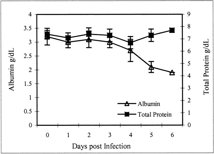
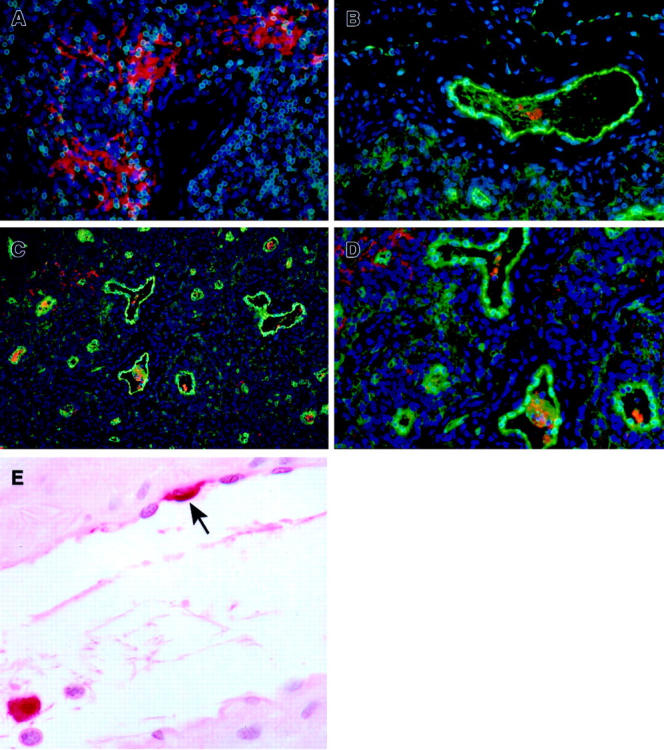
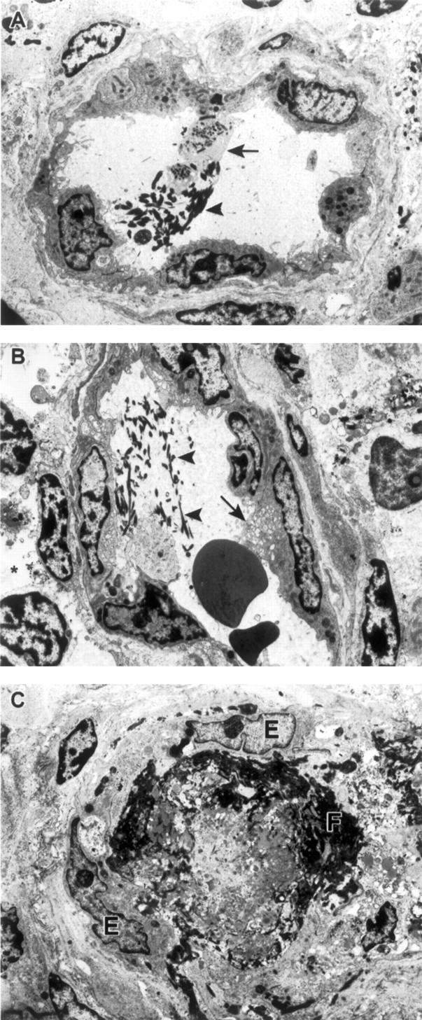
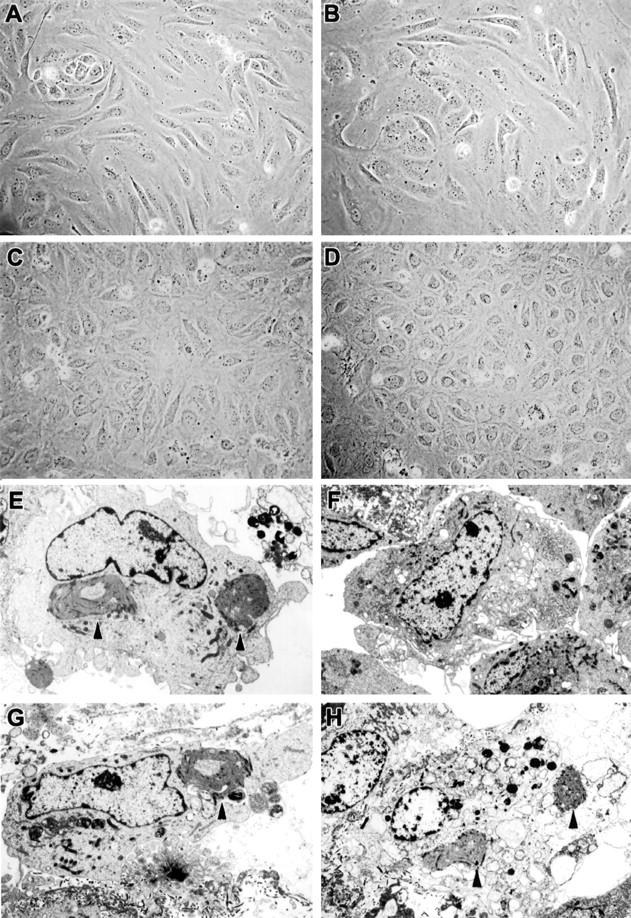
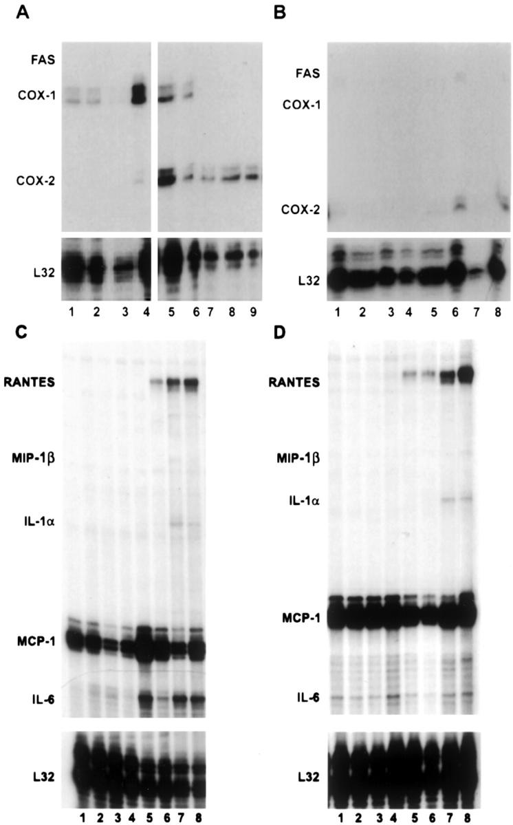
Ebola virus (EBOV) infection causes a severe and often fatal hemorrhagic disease in humans and nonhuman primates. Whether infection of endothelial cells is central to the pathogenesis of EBOV hemorrhagic fever (HF) remains unknown. To clarify the role of endothelial cells in EBOV HF, we examined tissues of 21 EBOV-infected cynomolgus monkeys throughout time, and also evaluated EBOV infection of primary human umbilical vein endothelial cells and primary human lung-derived microvascular endothelial cells in vitro. Results showed that endothelial cells were not early cellular targets of EBOV in vivo, as viral replication was not consistently observed until day 5 after infection, a full day after the onset of disseminated intravascular coagulation. Moreover, the endothelium remained relatively intact even at terminal stages of disease. Although human umbilical vein endothelial cells and human lung-derived microvascular endothelial cells were highly permissive to EBOV replication, significant cytopathic effects were not observed. Analysis of host cell gene response at 24 to 144 hours after infection showed some evidence of endothelial cell activation, but changes were unremarkable considering the extent of viral replication. Together, these data suggest that coagulation abnormalities associated with EBOV HF are not the direct result of EBOV-induced cytolysis of endothelial cells, and are likely triggered by immune-mediated mechanisms.


Figures

Figure 1.
Figure 1.
Total serum protein and albumin values after infection of cynomolgus monkeys with EBOV-Zaire. Note that day 0 values represent controls that were obtained before the animals were challenged with EBOV.
Figure 2.
Figure 2.
Immunohistochemistry of inguinal lymph nodes of EBOV-Zaire-infected cynomolgus monkeys for cell markers and EBOV at day 5. A: Abundance of EBOV-positive mononuclear cells (red) and extracellular EBOV antigen in extravascular area. Note absence of EBOV-positive endothelial cells. B–D: Double labeling for an endothelial cell marker (von Willebrand factor) (green) and EBOV antigens (red). Note: D is a higher magnification of C. E: Venule, brachial plexus. Rare EBOV-positive endothelial cell (arrow) and typical EBOV-positive monocyte (within lumen). A–D, Immunofluorescence method with 4′,6′-diamidino-2-phenylindole counterstain; E, alkaline phosphatase method. Original magnifications: ×20 (C); ×40 (A, B, D, E).
Figure 3.
Figure 3.
Ultrastuctural appearance of vessels in lymph nodes of EBOV-Zaire-infected cynomolgus monkeys. A: Intravascular fibrin deposits (arrowhead) and adherence of platelets (arrow) to vascular endothelium at day 4. B: Intravascular fibrin deposits (arrowheads), vacuolization of endothelial cell cytoplasm (arrow), and evidence of mild edema (asterisk) at day 4. C: Obstruction of vessel lumen with fibrin and fibrinocellar debris (F). Despite observed coagulopathy, endothelial cells (E) show no evidence of EBOV infection. Original magnifications, ×6500 (A–C).
Figure 4.
Figure 4.
EBOV replication in HUVECs and HMVEC-Ls by infectivity titration.
Figure 5.
Figure 5.
Cytopathology of EBOV-infected HUVECs and HMVEC-Ls at day 6. Inverted phase microscopy of mock-infected (A) and EBOV-infected (B) HUVECs, and mock-infected (C) and EBOV-infected (D) HMVEC-Ls. Note that there are relatively minor differences in cytopathology. E–H: Ultrastructural morphology. E: EBOV-infected HUVECs with typical EBOV intracytoplasmic inclusion (arrowheads). F: Mock-infected HMVEC-Ls. G: EBOV-infected HMVEC-Ls with typical EBOV intracytoplasmic inclusion (arrowhead). H: EBOV-infected HMVEC-Ls with typical EBOV intracytoplasmic inclusions (arrowheads) and morphological evidence of necrosis. Note comparable nuclear morphology in E, F, and G, whereas nuclei in H show degenerative necrotic change as evidenced by pale-staining and random clumping of chromatin. Original magnifications: ×4000 (E); ×3000 (F–H).
Figure 6.
Figure 6.
Analysis of mRNA production in EBOV-infected endothelial cell cultures. Representative RNase protection assays are shown. A: COX-1, COX-2, iNOS, Fas; comparison of mock-infected HUVECs at 1 hour (lane 1), 24 hours (lane 2), 96 hours, (lane 3), and 144 hours (lane 4) with EBOV-infected HUVECs at 1 hour (lane 5), 24 hours (lane 6), 96 hours (lane 7), and 144 hours (lane 8), and HUVECs treated with inactivated EBOV at 144 hours (lane 9). B: COX-1, COX-2, iNOS, Fas; comparison of mock-infected HMVEC-Ls at 1 hour (lane 1), 24 hours (lane 2), 96 hours (lane 3), and 144 hours (lane 4) with EBOV-infected HMVEC-Ls at 1 hour (lane 5), 24 hours (lane 6), 96 hours (lane 7), and 144 hours (lane 8). C: Cytokines/chemokines; comparison of mock-infected HUVECs at 1 hour (lane 1), 24 hours (lane 2), 96 hours (lane 3), and 144 hours (lane 4) with EBOV-infected HUVECs at 1 hour (lane 5), 24 hours (lane 6), 96 hours (lane 7), and 144 hours (lane 8). D: Cytokines/chemokines; comparison of mock-infected HMVEC-Ls at 1 hour (lane 1), 24 hours (lane 2), 96 hours (lane 3), and 144 hours (lane 4) with EBOV-infected HMVEC-Ls at 1 hour (lane 5), 24 hours (lane 6), 96 hours (lane 7), and 144 hours (lane 8).
Figure 7.
Figure 7.
Analysis of cytokine/chemokine and prostacyclin (measuring its stable metabolite, 6-keto-prostaglandin F) accumulation in culture fluids of EBOV-infected HUVECs and HMVEC-Ls by enzyme-linked immunosorbent assay.

References

    1. Huggins JW: Prospects for treatment of viral hemorrhagic fevers with ribavirin, a broad-spectrum antiviral drug. Rev Infect Dis Suppl 1989, 4:S750-S761 - PubMed
    1. Jahrling PB, Geisbert J, Swearengen JR, Jaax GP, Lewis T, Huggins JW, Schmidt JJ, LeDuc JW, Peters CJ: Passive immunization of Ebola virus-infected cynomolgus monkeys with immunoglobulin from hyperimmune horses. Arch Virol Suppl 1996, 11:135-140 - PubMed
    1. Jahrling PB, Geisbert TW, Geisbert JB, Swearengen JR, Bray M, Jaax NK, Huggins JW, LeDuc JW, Peters CJ: Evaluation of immune globulin and recombinant interferon-α2b for treatment of experimental Ebola virus infections. J Infect Dis Suppl 1999, 179:S224-S234 - PubMed
    1. Yang Z, Delgado R, Xu L, Todd RF, Nabel EG, Sanchez A, Nabel GJ: Distinct cellular interactions of secreted and transmembrane Ebola virus glycoproteins. Science 1998, 279:1034-1037 - PubMed
    1. Yang Z, Duckers HJ, Sullivan NJ, Sanchez A, Nabel EG, Nabel GJ: Identification of the Ebola virus glycoprotein as the main viral determinant of vascular cell cytotoxicity and injury. Nat Med 2000, 6:886-889 - PubMed

Publication types