. 2003 Dec;163(6):2503-12.
doi: 10.1016/s0002-9440(10)63605-x.

Expression profiling in ovarian clear cell carcinoma: identification of hepatocyte nuclear factor-1 beta as a molecular marker and a possible molecular target for therapy of ovarian clear cell carcinoma

Affiliations

Expression profiling in ovarian clear cell carcinoma: identification of hepatocyte nuclear factor-1 beta as a molecular marker and a possible molecular target for therapy of ovarian clear cell carcinoma

Akira Tsuchiya et al. Am J Pathol. 2003 Dec.

Abstract

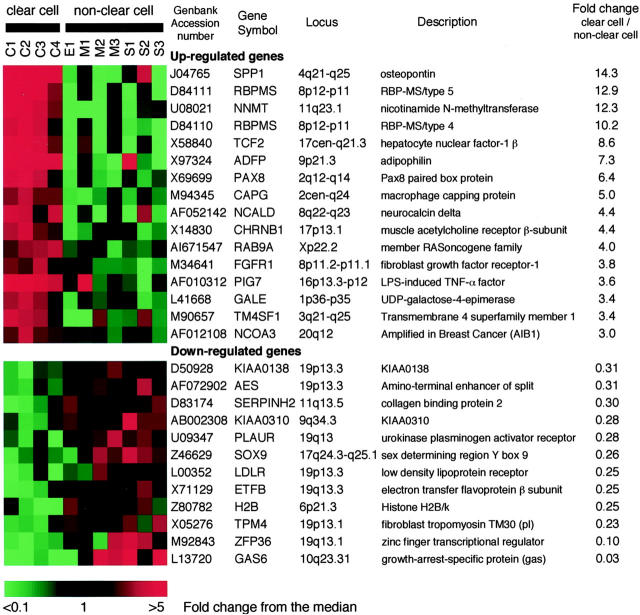
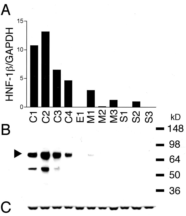
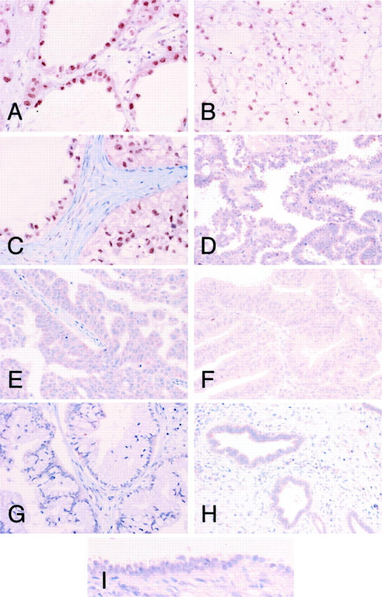
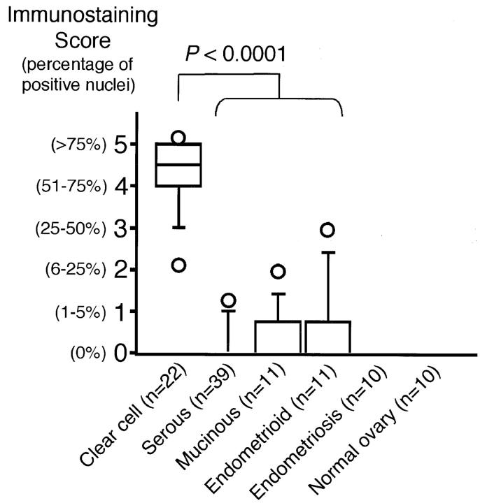
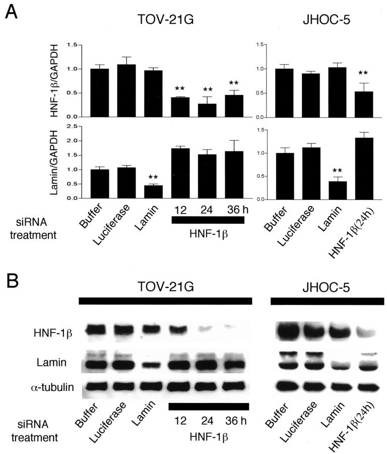
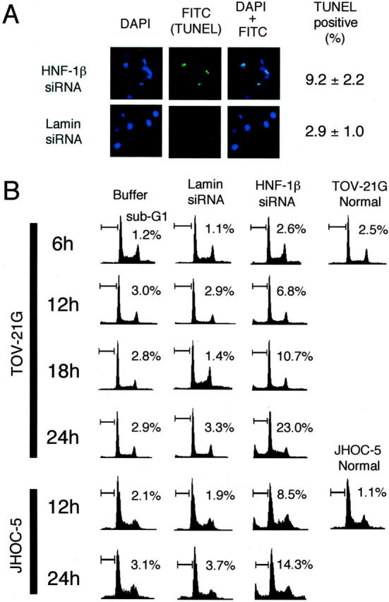
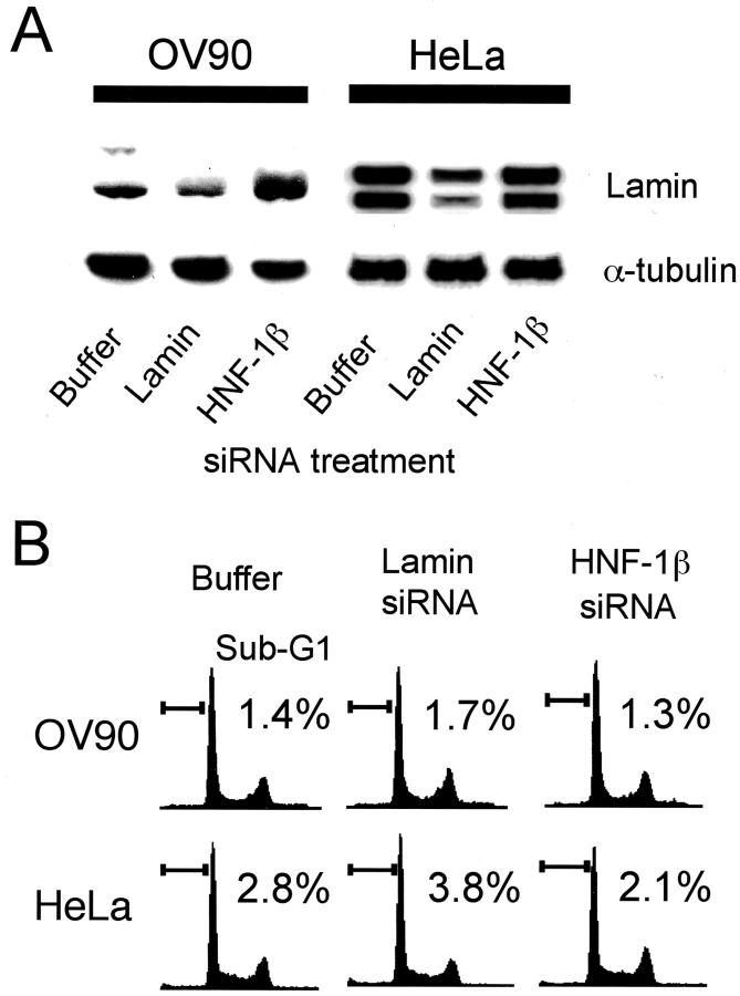
Of all of the epithelial ovarian cancers, clear cell carcinoma (CCC) of the ovary has the worst prognosis. We applied the oligonucleotide array technique to identify genes generally involved in CCC. Of the approximately 12,600 genes that were analyzed, 28 were expressed significantly differently between four CCC and seven non-CCC cell lines. Among 16 up-regulated genes in CCC, we further investigated a transcription factor, hepatocyte nuclear factor-1 beta (HNF-1 beta). We validated up-regulation of HNF-1 beta in CCC in terms of both mRNA and protein level using real-time quantitative reverse transcriptase-polymerase chain reaction and immunoblotting. Immunohistochemical analysis of 83 surgically resected ovarian cancers showed that almost all CCC specimens (21 of 22 cases) had nuclear staining for HNF-1 beta, whereas most non-CCC specimens (60 of 61 cases) showed no immunostaining or only focal and faint staining in the nucleus. Furthermore, we investigated the significance of HNF-1 beta expression in CCC using RNA interference. The reduction of HNF-1 beta expression by RNA interference induced apoptotic cell death in ovarian CCC cells, which was confirmed by terminal dUTP nick-end labeling and fluorescence-activated cell-sorting analyses. Our results suggest that HNF-1 beta is not only an excellent CCC-specific molecular marker but also a molecular target for therapy of ovarian CCC.


Figures

Figure 1.
Figure 1.
Genes differently expressed in clear cell versus non-CCC. Twenty-eight genes displayed a threefold or more increase or decrease in expression level that was significant at the P < 0.05 level by U-test. Each color patch in the resulting visual map represents the expression level of the associated gene in the cell line sample, with a continuum of expression levels from green (lowest) to bright red (highest). GenBank accession numbers, gene symbols, loci, gene descriptions, and the fold changes are shown. The scale bar reflects the fold increase (red) or decrease (green) for any given gene relative to the median level of expression across all samples. Samples are labeled as follows: C, clear cell; E, endometrioid; M, mucinous; S, serous carcinoma of the ovary.
Figure 2.
Figure 2.
Real-time quantitative RT-PCR analysis and immunoblot analysis of HNF-1β in ovarian cancer cell lines. A: Gene expression levels for HNF-1β. The expression levels were normalized with GAPDH mRNA in each sample. B and C: Immunoblot analysis of HNF-1β protein (B). Each cell line lysate (20 μg) was analyzed using anti-HNF-1β antibody. Arrowheads indicate the normal HNF-1β isoform. The blot was stripped and reprobed with anti-α-tubulin antibody to check for equal loading of total protein (C). Abbreviations used for these cell lines (C1, C2, C3, C4, E1, M1, M2, M3, S1, S2, and S3) and the cell line histologies are shown in Table 1 ▶ .
Figure 3.
Figure 3.
Immunohistochemical analysis of HNF-1β protein expression in surgically resected epithelial ovarian cancers. Immunohistochemistry on paraffin-embedded samples of clear cell (A, case 26; B, case 8; C, case 15), serous (D, case 35; E, case 59), endometrioid (F, case 18), and mucinous (G, case 81) adenocarcinomas of the ovary was performed with an antibody against HNF-1β. Ovarian endometriosis (H) and normal ovarian surface epithelium (I) were also immunostained with the same antibody. CCCs had strong nuclear staining for HNF-1β. Original magnifications: ×200 (A–H); ×400 (I).
Figure 4.
Figure 4.
Relationship between immunohistochemical expression of HNF-1β and histology. Box plot showing the relationship between HNF-1β immunostaining score and histology. The box encompasses the 25th through 75th percentiles of results obtained with the 50th percentile (median). The 5th and 95th percentiles are shown as Ο below and above the 10th and 90th percentile whisker caps, respectively. The immunostaining scores for CCCs were significantly higher than those for other histologies (P < 0.0001 by U-test).
Figure 5.
Figure 5.
Effect of small interfering RNA (siRNA) transfection in TOV-21G and JHOC-5 ovarian CCC cells. A: Gene expression levels for lamin and HNF-1β measured by real-time quantitative RT-PCR analysis. Buffer (PBS plus transfection reagent)-, luciferase siRNA-, and lamin siRNA-transfected TOV-21G and JHOC-5 cells were analyzed 24 hours after the beginning of transfection. HNF-1β siRNA-transfected cells were analyzed 12, 24, and 36 hours after the beginning of transfection. The expression levels were normalized with GAPDH mRNA in each sample. The average of three independent experiments is shown, and error bars indicate SD. **, Significant reduction compared with buffer only (P < 0.001 by U-test). B: Immunoblot analysis of lamin and HNF-1β protein in transfected TOV-21G and JHOC-5 cells. Each cell line lysate (20 μg) was analyzed using anti-lamin and anti-HNF-1β antibodies. The blot was stripped and reprobed with anti-α-tubulin antibody to check for equal loading of total protein.
Figure 6.
Figure 6.
Induction of apoptosis by reduction of HNF-1β expression in TOV-21G and JHOC-5 ovarian CCC cells using RNAi. A: TOV-21G cells were transfected with HNF-1β or lamin siRNA. Apoptosis was detected by the TUNEL method with FITC emission 24 hours after the beginning of transfection. The percentage of TUNEL-positive values corresponds to the average and SD of 10 determinations of slides from three independent experiments with three fields/slide. B: Apoptotic cells were evaluated by FACS analysis at 6, 12, 18, and 24 hours after the beginning of transfection with buffer (PBS plus transfection reagent), lamin siRNA, and HNF-1β siRNA. Values indicate the percentage of cells with a sub-G1 DNA content. Three experiments were performed in triplicate.
Figure 7.
Figure 7.
HNF-1β siRNA does not induce apoptosis in HeLa or ovarian serous adenocarcinoma cells. A: Immunoblot analysis of lamin protein in transfected OV-90 and HeLa cells. Each cell line lysate (20 μg) was analyzed using anti-lamin antibody. The blot was stripped and reprobed with anti-α-tubulin antibody to check for equal loading of total protein. B: Apoptotic cells were evaluated by FACS analysis at 24 hours after the beginning of transfection with buffer (PBS plus transfection reagent), lamin siRNA, and HNF-1β siRNA. Values indicate the percentage of cells with a sub-G1 DNA content.

References

    1. Scully RE, Young RH, Clement PB: Rosai J eds. Tumors of the Ovary, Maldeveloped Gonads, Fallopian Tube, and Broad Ligament. 1999:pp 27-50 Armed Forces Institute of Pathology Washington DC
    1. Piver MS: Ovarian carcinoma. A decade of progress. Cancer 1984, 54:2706-2715 - PubMed
    1. Parmar MKB, Adams M, Balestrino M, Bertelsen K, Bonazzi C, Calvert H, Colombo N, Delaloye JF, Durando A, Guthrie D, Hagen B, Harper P, Mangioni C, Perren T, Poole C, Qian W, Rustin G, Sandercock J, Tumolo S, Torri V, Vecchione F, Tinazzi A, Uscinska B, Collins S, Flann M, Buda A, Taylor B, Tannock I, Souhami R, Granzia-Valsecchi M: Paclitaxel plus carboplatin versus standard chemotherapy with either single-agent carboplatin or cyclophosphamide, doxorubicin, and cisplatin in women with ovarian cancer: the ICON3 randomised trial. Lancet 2002, 360:505-515 - PubMed
    1. Tammela J, Geisler JP, Eskew PN, Jr, Geisler HE: Clear cell carcinoma of the ovary: poor prognosis compared to serous carcinoma. Eur J Gynaecol Oncol 1998, 19:438-440 - PubMed
    1. O’Brien ME, Schofield JB, Tan S, Fryatt I, Fisher C, Wiltshaw E: Clear cell epithelial ovarian cancer (mesonephroid): bad prognosis only in early stages. Gynecol Oncol 1993, 49:250-254 - PubMed

Publication types

MeSH terms