. 2003 Dec 1;17(23):2902-21.
doi: 10.1101/gad.1144403. Epub 2003 Nov 21.

Hierarchical assembly of the budding yeast kinetochore from multiple subcomplexes

Affiliations

Hierarchical assembly of the budding yeast kinetochore from multiple subcomplexes

Peter De Wulf et al. Genes Dev. .

Abstract

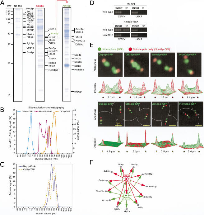
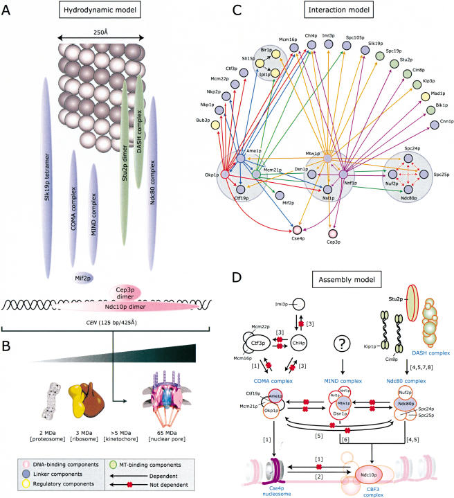
Kinetochores are multiprotein complexes that assemble on centromeric DNA and attach chromosomes to spindle microtubules. Over the past six years, the number of proteins known to localize to the Saccharomyces cerevisiae kinetochore has increased from around 10 to over 60. However, relatively little is known about the protein-protein interactions that mediate kinetochore assembly or about the overall structure of microtubule-attachment sites. Here we used biophysical techniques, affinity purification, mass spectrometry, and in vivo assays to examine the state of association of 31 centromere-binding proteins, including six proteins newly identified as kinetochore subunits. We found that yeast kinetochores resemble transcriptional enhancers in being composed of at least 17 discrete subcomplexes that assemble on DNA to form a very large structure with a mass in excess of 5 MD. Critical to kinetochore assembly are proteins that bridge subunits in direct contact with DNA and subunits bound to microtubules. We show that two newly identified kinetochore complexes, COMA (Ctf19p-Okp1p-Mcm21p-Ame1p) and MIND (Mtw1p including Nnf1p-Nsl1p-Dsn1p) function as bridges. COMA, MIND, and the previously described Ndc80 complex constitute three independent and essential platforms onto which outer kinetochore proteins assemble. In addition, we propose that the three complexes have different functions with respect to force generation and MT attachment.


Figures

Figure 1.
Figure 1.
Hydrodynamic analysis of kinetochore proteins. Whole-cell extracts were prepared from strains expressing either Slk19p-TAP or Stu2p-TAP (A,B), Ndc80p-6HA, Nuf2p-6HA, Spc24p-3HA or Spc25p-3HA, Dam1p-3Myc and Spc19p-ProA, Dam1p-3Myc and Spc34p-ProA (C,D), Mcm21p-TAP, Ctf19p-TAP, Okp1p-TAP, or Cse4p-TAP (E,F), and Mtw1p-ProA (G,H). Equal amounts of each extract were analyzed on a HiPrep 16/60 Sephacryl S-500 HR size-exclusion chromatography column and 5%-40% glycerol-density gradients. Fractions were immunoblotted with antisera that recognize the HA, Myc, TAP, or ProA epitope tags. Mif2p (A,B) and Ndc10p and Cep3p (E,F) were detected with polyclonal antibodies raised against recombinant protein.
Figure 2.
Figure 2.
Purification and identification of kinetochore proteins associated with Okp1p and Ame1p. (A) Left lane shows a colloidal blue-stained SDS PAGE gel of a mock immune purification from a wild-type strain (no tag). The labeled proteins are commonly found as contaminants in single-step affinity purifications from yeast. The center and right lanes are purifications from the Okp1p-3FLAG and Ame1p-3FLAG strains. Proteins that were highly enriched and absent in the control are labeled. The positions of some proteins differ from the expected molecular weights (Suppl. Table 2) as a consequence of protein degradation. (B) Hydrodynamic analysis of Ctf19p-TAP on size-exclusion chromatography compared to Cse4p-TAP, Mcm22p-ProA, and (C) Nkp1p-ProA. (D) Crosslinking of Ame1p-ProA to CENIV DNA in wild-type or ndc10-1 cells at 37°C using ChIP. DNA in immune complexes (IP) was amplified with primers to CENIV, or as negative controls, TELVI-R and URA3, and compared to DNA in whole-cell extracts (labeled “1% INPUT”). (E) Images of metaphase (upper panels) and anaphase (lower panels) cells carrying Spc42p-CFP (red) and Okp1p-GFP, Ctf19p-GFP, Ame1p-GFP, or Mcm21p-GFP (green). Images represent projections of 3D image stacks containing 10 to 20 0.2-μm sections and collected on a DeltaVision deconvolution microscope. The graphs show the intensity of the CFP (red) and GFP (green) signals along the axis of the spindle in arbitrary units. (F) Network summarizing interactions found among proteins present in Okp1p and Ame1p immune purifications (see Suppl. Table 2 for further information).
Figure 3.
Figure 3.
Ctf19p, Okp1p, Mcm21p, and Ame1p are components of the COMA complex. (A) Co-elution of Ctf19p-TAP, Okp1p-TAP, Mcm21p-TAP, and Ame1p-TAP by high-resolution size-exclusion chromatography. (B) Model for the predicted organization of Ame1p, Ctf19p, Mcm21p, and Okp1p subunits in the C1 and C2 COMA complexes based on the MW of the proteins: Ctf19p, 42 kD; Okp1p, 47 kD; Mcm21p, 37 kD; Ame1p, 37 kD; TAP tag, 20 kD. (C) Glycerol-density gradient analysis of Ame1p-TAP in okp1-5 cells at the permissive (24°C) and restrictive (37°C) temperatures. (D) Immunoblots using anti-ProA antibodies to detect the Ctf19p-ProA, Mcm21p-ProA, and Ame1p-TAP proteins in okp1-5 cells grown at 24°C or 37°C. The same extracts were probed with an anti-tubulin antibody as a loading control. The Nkp1p-ProA is included as a negative control. (E) Localization of Ame1p-GFP (green) in okp1-5 cells at 24°C and 37°C. (F) Glycerol-density gradient analysis of Ame1p-TAP in mtw1-1 cells grown at 24°C or 37°C and of Ame1p-TAP in (G) wild-type, ctf19Δ, mcm21Δ, and (H) mcm21Δctf19Δ cells. (I) Glycerol-density gradient analysis of Ame1p-TAP in cycling and α-factor arrested cells. (J) Localization of Ame1p-GFP (green) in wild-type, ctf19Δ, mcm21Δ, and mcm21Δctf19Δ cells.
Figure 4.
Figure 4.
Purification and identification of kinetochore proteins associated with Mtw1p. (A) Purification of Mtw1p-3FLAG, Nnf1p-3FLAG, and Mcm21p-3FLAG was carried out as described in the Figure 2A legend. (B) ChIP analysis and (C) localization of Nnf1p-GFP, Dsn1p-GFP, Nsl1p-GFP, Spc105p-GFP, and Cnn1p-GFP were as carried out as described in the Figure 2D,E legend. The expression levels of Cnn1p were very low, so we could not use Spc42p-CFP due to bleed-through of the CFP signal into the GFP channel. (D) Networks summarizing the interactions found among Mtw1p, Nnf1p, Mcm21p, and other kinetochore components. The solid lines depict the interactions identified by the proteins purifications shown in panel (A). The dashed lines show additional interactions identified in repeat purifications (see Suppl. Table 2 for further information).
Figure 5.
Figure 5.
The MIND complex contains Mtw1p, Nnf1p, Nsl1p, and Dsn1p. (A) Mtw1p-ProA, Dsn1p-ProA, Nsl1p-ProA, and Nnf1p-ProA co-elute on high-resolution size-exclusion chromatography. (B) Model for the predicted organization of Mtw1p, Dsn1p, Nsl1p, and Nnf1p in the M1, M2, and M3subcomplexes based on the molecular weight of the individual proteins: Mtw1p, 33 kD; Dsn1p, 65 kD; Nsl1p, 25 kD; Nnf1p, 24 kD; ProA tag, 15 kD. (C) Anti-ProA immunoblots of nnf1-17 cells carrying Nsl1p-ProA, Dsn1p-ProA, or Mtw1p-ProA grown at 24°C or 37°C. (D) Crosslinking of Nsl1p-ProA and Dsn1p-ProA to CEN DNA in nnf1-17 cells grown at 24°C or 37°C. (E) Anti-ProA immunoblots of whole-cell extracts, and (F) crosslinking of Nsl1p-ProA, Dsn1p-ProA, and Nnf1p-ProA to CEN DNA in mtw1-1 cells grown at 24°C or 37°C. (G) As a control, extracts were prepared from Mtw1p-ProA okp1-5 cells grown at 24°C or 37°C and analyzed by glycerol-density gradient ultracentrifugation.
Figure 6.
Figure 6.
Assembly of the COMA, MIND, and Ndc80 complexes at kinetochores. (A) Crosslinking of Ame1p-GFP to CEN DNA in wild-type, okp1-5, ndc80-1, and mtw1-1 cells grown at 37°C. (B) Localization of Ame1p-GFP (green) in wild-type, okp1-5, ndc80-1, and mtw1-1 cells grown at 37°C. (C) Crosslinking of Mtw1p-ProA to CEN DNA in spc25-7 cells grown at 24°C or 37°C and in wild-type, okp1-5, and nnf1-17 cells grown at 37°C. Ame1p-ProA acts as the positive control. (D) Crosslinking of Ndc80p-GFP to CEN DNA in wild-type, okp1-5, and mtw1-1 cells grown at 37°C. Crosslinking of Cin8p-GFP to CEN DNA in wild-type and ndc80-1 cells grown at 37°C (negative control).
Figure 7.
Figure 7.
Functional analysis of the COMA and MIND complexes in chromosome segregation. (A) Strains carrying a centromere-proximal chrIV tag and Spc42p-GFP (He et al. 2000) were synchronized with α-factor and released into prewarmed media at 37°C. Cells were fixed after 75 min, and the extent of transient sister separation was determined in metaphase cells (as determined by nuclear morphology and spindle length). Data for ndc80-1 are provided for comparison (He et al. 2001). (B) okp1-5 and mtw1-1 cells containing Ndc80p-GFP (green) and ndc80-1 cells containing Mtw1p-GFP (green) were shifted to 37°C for 3 h, fixed, and imaged as described in the Figure 2E legend. All strains contained Spc42p-CFP (red) to mark the position of the SPBs, which are indicated by the red arrows. (C) Schematic of the chrIV tag and Spc42p-GFP SPB tag showing the spindle-centered cylindrical reference system and three key parameters used to characterize chromosome motion: d1, the distance from the reference spindle pole to one chromatid tag; d2, the distance from the reference spindle pole to the sister chromatid; and d3, the distance between the SPBs. (D-O, left column) Scatter plots of the positions of chromosome tags over 300 sec in a time-lapse movie in wild-type, ndc80-1, okp1-5, and mtw1-1 cells at 37°C. The length of the spindle is shown in red; in general, spindles are longer in cells with kinetochore mutants than in wild-type cells, as a consequence of reduced inward directed spindle forces (Goshima and Yanagida 2000). Separate green and black lines are seen only when chromatids are transiently separated. The positions of chromosomes are viewed along the spindle axis. (D-O, middle column). Scatter plots with the spindle axis rotated -45° and elevated 20°. (D-O, right column) Plots of spindle length, d3 in part C (red), the distance between a reference SPB and the first chromosome tag, d1 (black) and between the reference SPB and the second chromosome tag, d2 (green). Distinct black and green lines are seen only when sister chromatids are undergoing transient separation.
Figure 8.
Figure 8.
Models of the structure, organization, and assembly of budding yeast kinetochores. (A) Hydrodynamic data from Table 1 were used to draw 3D ellipsoids for each kinetochore complex such that the volume was proportional to the molecular weight, and the axial ratio is represented as the degree of elongation of the ellipse. The single MT and CEN DNA are drawn approximately to scale. (B) Predicted size of the yeast kinetochore assuming that each known subunit is present in a single copy unless the subunit is known to be a homomultimer. (C) Protein-interaction model summarizing interactions among Okp1p (solid red arrows), Ame1p (solid blue arrows), Mcm21p (solid green arrows), Mtw1p (solid orange arrows), and Nnf1p (solid purple arrows). Interactions among Ndc80p complex subunits (dotted red arrows) are described by Wigge and Kilmartin (2001) and Janke et al. (2001). Interactions found with Sli15p (dotted black arrows) are described by Cheeseman et al. (2002b). (D) Assembly model for the COMA, MIND, and Ndc80 complexes at kinetochores. Black arrows represent dependencies (CEN binding by Mtw1p requires NDC10, and the arrow therefore connects the MIND and CBF3 complexes); a red “X” represents a lack of dependency. (For references see: [1] Measday et al. 2002, [2] Ortiz et al. 1999, [3] Pot et al. 2003, [4] He et al. 2001, [5] Janke et al. 2001, [6] Goshima and Yanagida 2000, [7] Janke et al. 2002, [8] J. Tytell and P. Sorger, unpubl.)

Similar articles

Cited by

References

    1. Bader G.D., Heilbut, A., Andrews, B., Tyers, M., Hughes, T., and Boone, C. 2003. Functional genomics and proteomics: Charting a multidimensional map of the yeast cell. Trends Cell. Biol. 13: 344-356. - PubMed
    1. Bell S.P. and Dutta, A. 2002. DNA replication in eukaryotic cells. Annu. Rev. Biochem. 71: 333-374. - PubMed
    1. Cheeseman I.M., Brew, C., Wolyniak, M., Desai, A., Anderson, S., Muster, N., Yates, J.R., Huffaker, T.C., Drubin, D.G., and Barnes, G. 2001. Implication of a novel multiprotein Dam1p complex in outer kinetochore function. J. Cell Biol. 155: 1137-1145. - PMC - PubMed
    1. Cheeseman I.M., Drubin, D.G., and Barnes, G. 2002a. Simple centromere, complex kinetochore: Linking spindle microtubules and centromeric DNA in budding yeast. J. Cell Biol. 157: 199-203. - PMC - PubMed
    1. Cheeseman I.M., Anderson, S., Jwa, M., Green, E.M., Kang, J., Yates 3rd, J.R., Chan, C.S., Drubin, D.G., and Barnes, G. 2002b. Phospho-regulation of kinetochore-microtubule attachments by the Aurora kinase Ipl1p. Cell 111: 163-172. - PubMed

Publication types

MeSH terms

Substances