. 2003 Dec 1;17(23):2922-32.
doi: 10.1101/gad.1156903. Epub 2003 Nov 21.

DNA damage response and MCL-1 destruction initiate apoptosis in adenovirus-infected cells

Affiliations

DNA damage response and MCL-1 destruction initiate apoptosis in adenovirus-infected cells

Andrea Cuconati et al. Genes Dev. .

Abstract

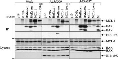
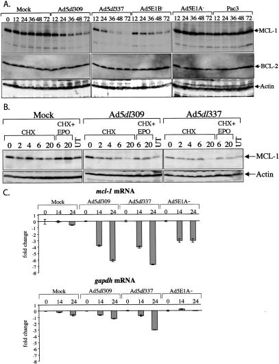
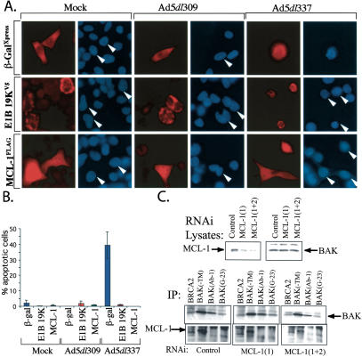
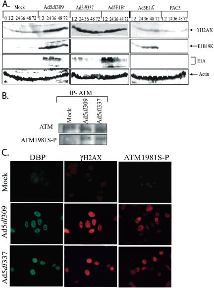
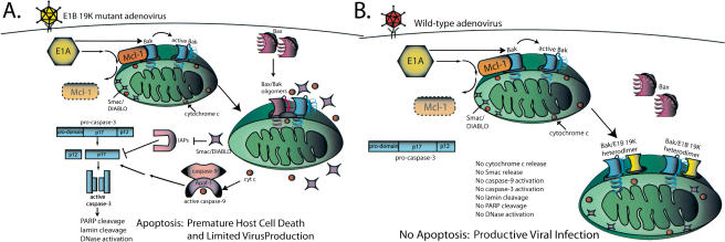
Expression of adenovirus E1A deregulates cell proliferation to facilitate viral DNA replication, prompting the initiation of apoptosis signaled primarily through proapoptotic BAK in productively infected cells. We demonstrate here that in uninfected cells, BAK is complexed with the anti-apoptotic BCL-2 family member Myeloid Cell Leukemia 1 (MCL-1). E1A expression during infection resulted in the specific down-regulation of MCL-1 through destabilization of the protein and loss of the mRNA. Upon loss of the MCL-1-BAK complex, BAK complexed with either BAX in proapoptotic E1B mutant adenovirus-infected cells, or with the adenovirus BCL-2 homolog E1B 19K in cells infected with the wild-type virus in which apoptosis is inhibited. Loss of MCL-1 was required to initiate the apoptotic pathway in infected cells as restoration of MCL-1 expression rescued infected cells from E1A-induced apoptosis. Analogous to E1A expression, DNA damage down-regulates MCL-1, and adenovirus infection resulted in the accumulation of phosphorylated H2AX and ataxia-telangiectasia mutant protein (ATM), hallmarks of DNA double-strand breaks. Thus, MCL-1 may function by maintaining BAK in an inactive state, and the loss of MCL-1 upon activation of the DNA damage response, perhaps through replication stress induced in virus infected cells, may be required to initiate the apoptotic response.


Figures

Figure 1.
Figure 1.
BAK and MCL-1 form a complex that is disrupted during adenovirus infection. Immunoprecipitation (IP) of BAK and MCL-1 from mock, Ad5dl309, Ad5dl337 infected cells was carried out with anti-p19, anti-PCNA, anti-BAK(Ab-1), anti-BAK(TM), anti-MCL-1, anti-MCL-1(Ab-1) antibodies (Abs) from the soluble fraction of cells lysed in CHAPS-containing buffer at 24 h post-infection. Mock-infected HeLa cell lysate (M) was utilized as a marker for MCL-1, BAK, and BAX expression levels. Western blotting was carried out on precipitated material with an anti-MCL-1 antibody, anti-BAK plus anti-BAX antibodies, or an anti-E1B 19K antibody. Samples of lysates collected before IP (Lysates) were analyzed to ascertain total protein levels prior to immunoprecipitation.
Figure 2.
Figure 2.
E1A expression during adenovirus infection causes the loss of MCL-1 protein. (A) HeLa cells were mock, Ad5dl309, Ad5dl337, Ad5E1B-, Ad5E1A-, or Pac3 infected, and representative samples were collected at 0 (for mock only), 12, 24, 36, 48, and 72 h post-infection, and analyzed by Western blotting with antibodies against MCL-1, BCL-2, and actin. (B) Adenovirus infection stimulates proteasome-mediated turnover of MCL-1. HeLa cells were mock, Ad5dl309, or Ad5dl337 infected for 24 h, and then treated with cycloheximide (CHX) for 0, 2, 4, 6, and 20 h, or a combination of CHX plus epoxomicin (EPO) for 6 or 20 h, or were left untreated (UT). Samples were collected at the indicated time points and analyzed by Western blotting with anti-MCL-1 and anti-actin antibodies. (C) The effect of adenovirus infection on mcl-1 mRNA levels. Total RNA from mock, Ad5dl309, Ad5dl337, or Ad5E1A- infected cells was isolated at 0, 14, and 24 h post-infection, and was analyzed by Taqman real-time PCR using a probe and primers specific for mcl-1 mRNA. Fold change represents the difference in fluorescence signal in the 14- and 24-h samples compared with the corresponding 0-h sample. Error bars, the standard deviation of triplicate samples. Samples were also analyzed by Taqman real-time PCR with primers and a probe specific for gapdh.
Figure 3.
Figure 3.
Exogenous MCL-1 overexpression rescues apoptosis in adenovirus-infected cells. (A) MCL-1 prevents apoptosis-associated chromatin condensation in Ad5dl337-infected cells. HeLa cells expressing FLAG-MCL-1, E1B 19K-V5, and β-Gal-Xpress by transfection were mock, Ad5dl309, or Ad5dl337 infected for 30 h, and analyzed by immunofluorescence staining and Hoescht's dye DNA staining for chromatin condensation. Rhoadmine (red) stain represents cells expressing the transfected gene, whereas blue stain represents chromatin staining. White arrows indicate the corresponding chromatin-stained cells from the matching rhodamine-stained micrograph. (B) MCL-1 expression rescues infected cells from adenovirus-induced apoptosis in the absence of E1B 19K. The percentage of transfected mock, Ad5dl309, or Ad5dl337 infected cells exhibiting chromatin condensation by Hoescht's dye staining was determined by counting only those staining positive for the transfected gene. Error bars, the standard deviation of counts from two separate experiments. (C) Down-regulation of MCL-1 is not sufficient for BAK activation. HeLa cells were transfected with either control or mcl-1-specific siRNAs (1) or (1+2). Levels of MCL-1 and BAK were determined by Western blotting (top) and BAK activation was determined by IP with BAK antibodies (bottom). Enhanced BAK (Ab-1) immunoprecipitation indicative of BAK activation occurs in apoptotic cells (see Fig. 1). BAK-MCL-1 coimmunoprecipitation with the BAK-TM antibody is observed in the MCL-1 Western blot of the BAK immunoprecipitations (bottom).
Figure 4.
Figure 4.
Induction of H2AX and ATM phosphorylation during virus infection. (A) HeLa cells were mock, Ad5dl309, Ad5dl337, Ad5E1B-, Ad5E1A-, or Pac3 infected, and representative samples were collected at 0 (for mock only), 12, 24, 36, 48, and 72 h post-infection (see Fig. 2) and analyzed by Western blotting with antibodies against γH2AX (phospho-serine 139-specific form of H2AX), E1B 19K, E1A, and actin. (B) ATM was immunoprecipitated from Mock, Ad5dl309, and Ad5dl337 infected HeLa cells (MOI 100 at 24 h post-infection) and was Western blotted for either ATM or ATM1981S-P (phospho-serine 1981-specific form of ATM), as indicated. (C) Mock, Ad5dl309, and Ad5dl337 infected HeLa cells (MOI 100 at 24 h post-infection) were fixed and stained for indirect immunoflourescence with antibodies directed against the adenovirus 72 kD DBP, γH2AX (double-label indirect immunflourescence), and ATM1981S-P, as indicated.
Figure 5.
Figure 5.
Schematic representation of the regulation of adenovirus-induced apoptosis in infected cells. See text for explanation.

References

    1. Bae J., Leo, C.P., Hsu, S.Y., and Hsuch, A.J. 2000. MCL-1S, a splicing variant of the antiapoptotic BCL-2 family member MCL-1, encodes a proapoptotic protein possessing only the BH3 domain. J. Biol. Chem. 275: 25255-25261. - PubMed
    1. Bakkenist C. and Kastan, M.B. 2003. DNA damage activates ATM through intermolecular autophosphorylation and dimer dissociation. Nature 421: 499-506. - PubMed
    1. Bassing C.H., Suh, H., Ferguson, D.O., Chua, K.F., Manis, J., Eckersdorff, M., Gleason, M., Bronson, R., Lee, C., and Alt, F.W. 2003. Histone H2AX: A dosage-dependent suppressor of oncogenic translocations and tumors. Cell 114: 359-370. - PubMed
    1. Brader K.R., Wolf, J.K., Hung, M.C., Yu, D., Crispens, M.A., van Golen, K.L., and Price, J.E. 1997. Adenovirus E1A expression enhances the sensitivity of an ovarian cancer cell line to multiple cytotoxic agents through an apoptotic mechanism. Clin. Cancer Res. 3: 2017-2024. - PubMed
    1. Celeste A., Diflippantonio, S., Diflippantonio, M.J., Fernandez-Capetillo, O., Pilch, D.R., Sedelnikova, O.A., Eckhaus, M., Ried, T., Bonner, W.M., and Nussenzweig, A. 2003. H2AX haploinsufficiency modifies genomic stability and tumor susceptibility. Cell 114: 371-383. - PMC - PubMed

Publication types