

Three-dimensional structure of the bacterial multidrug transporter EmrE shows it is an asymmetric homodimer
- PMID: 14633977
- PMCID: PMC291852
- DOI: 10.1093/emboj/cdg611
Three-dimensional structure of the bacterial multidrug transporter EmrE shows it is an asymmetric homodimer
Abstract
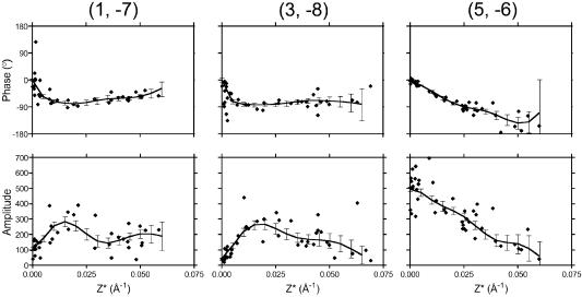
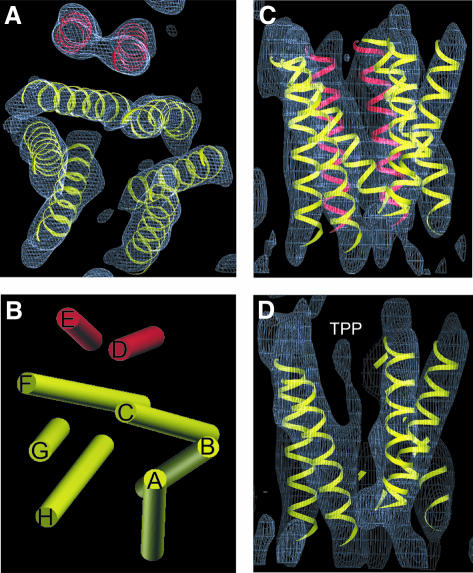
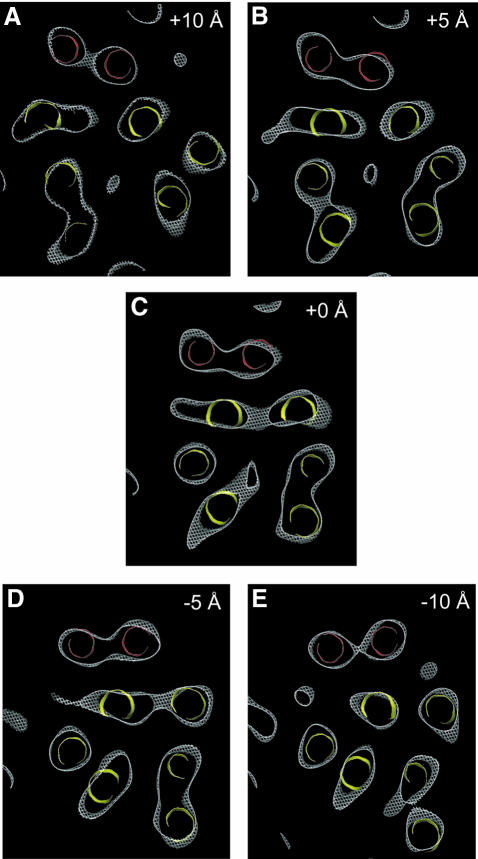
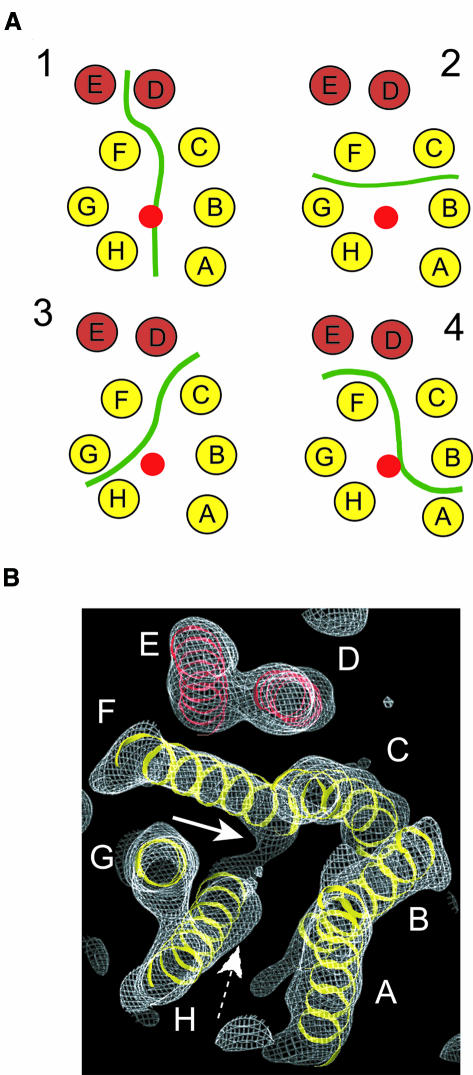
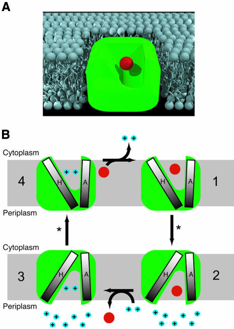
The small multidrug resistance family of transporters is widespread in bacteria and is responsible for bacterial resistance to toxic aromatic cations by proton-linked efflux. We have determined the three-dimensional (3D) structure of the Escherichia coli multidrug transporter EmrE by electron cryomicroscopy of 2D crystals, including data to 7.0 A resolution. The structure of EmrE consists of a bundle of eight transmembrane alpha-helices with one substrate molecule bound near the centre. The substrate binding chamber is formed from six helices and is accessible both from the aqueous phase and laterally from the lipid bilayer. The most remarkable feature of the structure of EmrE is that it is an asymmetric homodimer. The possible arrangement of the two polypeptides in the EmrE dimer is discussed based on the 3D density map.
Figures

Similar articles
-
New insights into the structure and oligomeric state of the bacterial multidrug transporter EmrE: an unusual asymmetric homo-dimer.FEBS Lett. 2004 Apr 30;564(3):234-8. doi: 10.1016/S0014-5793(04)00228-5. FEBS Lett. 2004. PMID: 15111102
-
X-ray structure of the EmrE multidrug transporter in complex with a substrate.Science. 2005 Dec 23;310(5756):1950-3. doi: 10.1126/science.1119776. Science. 2005. Retraction in: Science. 2006 Dec 22;314(5807):1875. doi: 10.1126/science.314.5807.1875b. PMID: 16373573 Retracted.
-
The projection structure of EmrE, a proton-linked multidrug transporter from Escherichia coli, at 7 A resolution.EMBO J. 2001 Jan 15;20(1-2):77-81. doi: 10.1093/emboj/20.1.77. EMBO J. 2001. PMID: 11226157 Free PMC article.
-
Comparison of three structures of the multidrug transporter EmrE.Curr Opin Struct Biol. 2006 Aug;16(4):457-64. doi: 10.1016/j.sbi.2006.06.005. Epub 2006 Jul 7. Curr Opin Struct Biol. 2006. PMID: 16828280 Review.
-
Precious things come in little packages.J Mol Microbiol Biotechnol. 2001 Apr;3(2):155-62. J Mol Microbiol Biotechnol. 2001. PMID: 11321568 Review.
Cited by
-
Structural Insights into Transporter-Mediated Drug Resistance in Infectious Diseases.J Mol Biol. 2021 Aug 6;433(16):167005. doi: 10.1016/j.jmb.2021.167005. Epub 2021 Apr 20. J Mol Biol. 2021. PMID: 33891902 Free PMC article. Review.
-
pH-induced structural change in a sodium/proton antiporter from Methanococcus jannaschii.EMBO J. 2005 Aug 3;24(15):2720-9. doi: 10.1038/sj.emboj.7600727. Epub 2005 Jul 14. EMBO J. 2005. PMID: 16015376 Free PMC article.
-
A structural model of EmrE, a multi-drug transporter from Escherichia coli.Biophys J. 2004 Jun;86(6):3335-48. doi: 10.1529/biophysj.103.034546. Biophys J. 2004. PMID: 15189838 Free PMC article.
-
EmrE dimerization depends on membrane environment.Biochim Biophys Acta. 2014 Jul;1838(7):1817-22. doi: 10.1016/j.bbamem.2014.03.013. Epub 2014 Mar 26. Biochim Biophys Acta. 2014. PMID: 24680655 Free PMC article.
-
Protonation of a glutamate residue modulates the dynamics of the drug transporter EmrE.Nat Chem Biol. 2016 Mar;12(3):141-5. doi: 10.1038/nchembio.1999. Epub 2016 Jan 11. Nat Chem Biol. 2016. PMID: 26751516 Free PMC article.
References
-
- Abramson J., Smirnova,I., Kasho,V., Verner,G., Kaback,H.R. and Iwata,S. (2003) Structure and mechanism of the lactose permease of Escherichia coli. Science, 301, 610–615. - PubMed
-
- Arkin I.T., Russ,W.P., Lebendiker,M. and Schuldiner,S. (1996) Determining the secondary structure and orientation of EmrE, a multi-drug transporter, indicates a transmembrane four-helix bundle. Biochemistry, 35, 7233–7238. - PubMed
-
- Chang G. (2003) Structure of MsbA from Vibrio cholera: a multidrug resistance ABC transporter homolog in a closed conformation. J. Mol. Biol., 330, 419–430. - PubMed
-
- Chang G. and Roth,C.B. (2001) Structure of MsbA from E. coli: a homolog of the multidrug resistance ATP binding cassette (ABC) transporters. Science, 293, 1793–1800. - PubMed
-
- Cizman M. (2003) The use and resistance to antibiotics in the community. Int. J. Antimicrob. Agents, 21, 297–307. - PubMed
Publication types
MeSH terms
Substances
Grants and funding
LinkOut - more resources
Full Text Sources
Other Literature Sources
Molecular Biology Databases
