. 2003 Dec 1;22(23):6182-92.
doi: 10.1093/emboj/cdg608.

Structure of the mitochondrial ATP synthase by electron cryomicroscopy

Affiliations

Structure of the mitochondrial ATP synthase by electron cryomicroscopy

John L Rubinstein et al. EMBO J. .

Abstract

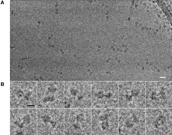
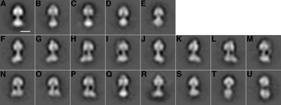
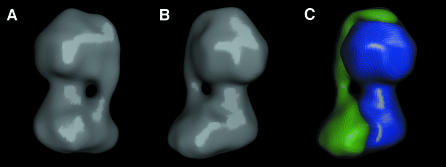
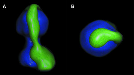
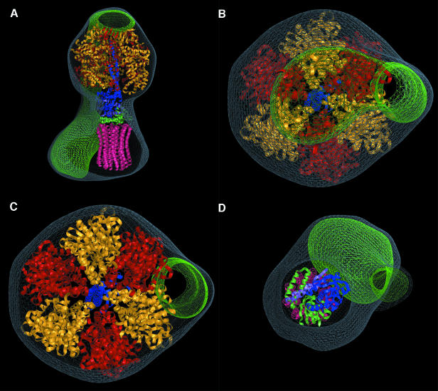
We have determined the structure of intact ATP synthase from bovine heart mitochondria by electron cryomicroscopy of single particles. Docking of an atomic model of the F1-c10 subcomplex into a major segment of the map has allowed the 32 A resolution density to be interpreted as the F1-ATPase, a central and a peripheral stalk and an FO membrane region that is composed of two domains. One domain of FO corresponds to the ring of c-subunits, and the other probably contains the a-subunit, the transmembrane portion of the b-subunit and the remaining integral membrane proteins of FO. The peripheral stalk wraps around the molecule and connects the apex of F1 to the second domain of FO. The interaction of the peripheral stalk with F1-c10 implies that it binds to a non-catalytic alpha-beta interface in F1 and its inclination where it is not attached to F1 suggests that it has a flexible region that can serve as a stator during both ATP synthesis and ATP hydrolysis.


Figures

None
Fig. 1. EM of ATP synthase particles. Frozen ATP synthase particles were imaged with a 200 kV electron microscope. (A) A sample region from a micrograph. (B) Some typical particle images. The scale bar in (A) represents 200 Å and the scale bar in (B) represents 100 Å.
None
Fig. 2. Class-average images of the ATP synthase. (A) Average of all of the aligned images in the dataset. The scale bar represents 100 Å. (BE) Class-averages that probably arise from incoherently averaged particle images (and consequently, like the overall average, have a line of symmetry). (FM) Class-averages where a corresponding mirror image is found (e.g. F with J, G with K, etc.) and constitute views about the long axis of the complex. (N and O) A mirror pair, but each probably consisting of averages of two non-equivalent views of the assembly with the long axis tilted from the plane of the grid. (PU) Class averages that have no mirror pair in the dataset.
None
Fig. 3. Rotation analysis. (A) Features that deviated from cylindrical symmetry in class-averages of Figure 2F–M were emphasized by subtracting the average of all of the views from each of the views. Markers were placed at maxima in the density to indicate the corresponding asymmetric features in each image. (B) To compare the experimental marker positions to the hypothesis that the images of the ATP synthase constitute a single-axis rotation series, the ratio of marker position to calculated marker radius (ai,j/ri) was plotted against the total angle for each marker in each image (φj + δi). The observations are consistent with the hypothesis that the series constitutes a rotation sequence, indicated by the dashed line (cos[φj + δi]).
None
Fig. 4. 3-D model of the ATP synthase. (A and B) Surface rendered views of the model after refinement. (C) The model after being divided into two parts. The first (blue) was chosen to correspond to the F1-c10 subcomplex and the remaining density (green) is interpreted to represent the peripheral stalk and second domain of FO. The grey mesh represents the experimental EM map.
None
Fig. 5. Two further views of the peripheral stalk. (A) A side-view of the model shows the curvature of the peripheral stalk. (B) A view from the F1 end of the assembly along its long axis reveals a sharp bend in the peripheral stalk near to its terminus.
None
Fig. 6. Determination of the absolute hand of the model. Tilt pairs of 29 different particles were recorded with a tilt between images of 30°. The first particle in each pair was aligned to the model. The plot shows the average phase residual between the model and the second particle in each pair for all possible tilt axes and tilts up to 40°. From the known direction and magnitude of the tilt between the images in the pair, a model of the correct hand was expected to produce a minimum at (2,–30°) as opposed to (–2,30°). The observed tilt axis and amount of tilt indicate that the model, as shown, is correct.
None
Fig. 7. Docking of an atomic model of F1-c10 into the EM map. The volume of the EM map not occupied by the atomic structure is coloured green. (A) A side-view of the model. The density that cannot be explained by the F1-c10 model consists of the peripheral stalk and the second domain of the FO region. This density must contain the remaining subunits of the ATP synthase: a, b, d, e, f, g, A6L, F6 and OSCP. (BD) Cross-sections of the model. The terminus of the peripheral stalk at the top of F1 is on the central axis at the apex of the model (B). The peripheral stalk is primarily in contact with an α-subunit (red) near the top of F1, but is in contact with a non-catalytic interface between an α- (red) and β- (yellow) subunit along most of F1. The matching flattened sides of the F1 in the crystal structure and EM map show the uniqueness of fit between the two models. The central stalk and c-ring fit into the model well (D).

Similar articles

Cited by

References

    1. Abrahams J.P., Leslie,A.G., Lutter,R. and Walker,J.E. (1994) Structure at 2.8 Å resolution of F1-ATPase from bovine heart mitochondria. Nature, 370, 621–628. - PubMed
    1. Bellare J.R., Davis H.T., Scriven,L.E. and Talmon,Y. (1988) Controlled environment vitrification system: an improved sample preparation technique. J. Electron Microsc. Tech., 10, 87–111. - PubMed
    1. Böttcher B., Schwarz,L. and Gräber,P. (1998) Direct indication for the existence of a double stalk in CFoF1. J. Mol. Biol., 281, 757–762. - PubMed
    1. Böttcher B., Bertsche,I., Reuter,R. and Gräber,P. (2000) Direct visualisation of conformational changes in EFOF1 by electron microscopy. J. Mol. Biol., 296, 449–457. - PubMed
    1. Boyer P.D. (1997) The ATP synthase—a splendid molecular machine. Annu. Rev. Biochem., 66, 717–749. - PubMed

MeSH terms

Substances