. 2003 Dec 1;22(23):6234-44.
doi: 10.1093/emboj/cdg596.

The phosphorylation state of an autoregulatory domain controls PACS-1-directed protein traffic

Affiliations

The phosphorylation state of an autoregulatory domain controls PACS-1-directed protein traffic

Gregory K Scott et al. EMBO J. .

Abstract

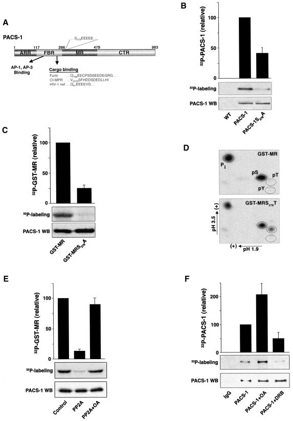
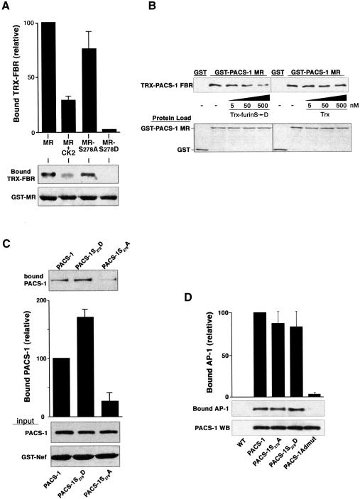
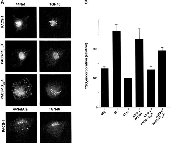
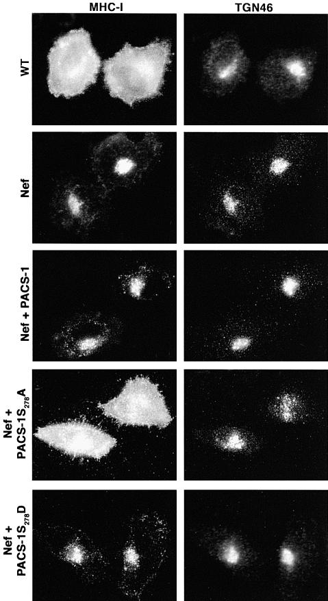
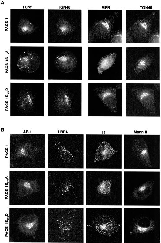
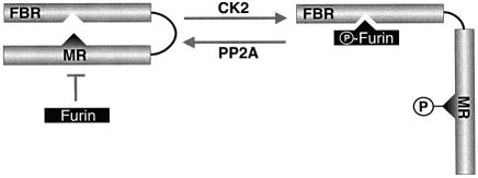
PACS-1 is a cytosolic sorting protein that directs the localization of membrane proteins in the trans-Golgi network (TGN)/endosomal system. PACS-1 connects the clathrin adaptor AP-1 to acidic cluster sorting motifs contained in the cytoplasmic domain of cargo proteins such as furin, the cation-independent mannose-6-phosphate receptor and in viral proteins such as human immunodeficiency virus type 1 Nef. Here we show that an acidic cluster on PACS-1, which is highly similar to acidic cluster sorting motifs on cargo molecules, acts as an autoregulatory domain that controls PACS-1-directed sorting. Biochemical studies show that Ser278 adjacent to the acidic cluster is phosphorylated by CK2 and dephosphorylated by PP2A. Phosphorylation of Ser278 by CK2 or a Ser278-->Asp mutation increased the interaction between PACS-1 and cargo, whereas a Ser278-->Ala substitution decreased this interaction. Moreover, the Ser278-->Ala mutation yields a dominant-negative PACS-1 molecule that selectively blocks retrieval of PACS-1-regulated cargo molecules to the TGN. These results suggest that coordinated signaling events regulate transport within the TGN/endosomal system through the phosphorylation state of both cargo and the sorting machinery.


Figures

None
Fig. 1. PACS-1 Ser278 is phosphorylated by CK2 and dephosphorylated by PP2A. (A) Schematic diagram of PACS-1 showing the atrophin-related region (ARR), the furin-binding region (FBR) which interacts with cargo molecules and AP-1/AP-3 adaptor complexes, the middle region (MR) which contains the autoregulatory acidic cluster and Ser278, and the C-terminal region (CTR). The acidic cluster in the PACS-1 MR as well as acidic cluster sequences contained in membrane cargo molecules, which bind to the PACS-1 FBR, are indicated. (BIn vivo phosphorylation of expressed PACS-1. BSC40 cells infected with VV recombinants expressing either epitope (HA)-tagged PACS-1 or PACS-1S278A were labeled with 32Pi, and the immunoprecipitated PACS-1 proteins were separated by SDS–PAGE and analyzed by autoradiography (upper panel). A western blot using the anti-HA mAb HA.11 shows equal expression and loading of the two proteins (lower panel). (CIn vitro CK2 phosphorylation of GST–MR. GST–MR or GST–MRS278A was incubated with CK2 and [γ-32P]ATP, separated by SDS–PAGE and analyzed by autoradiography. (D) PACS-1 MR is phosphorylated at amino acid 278. GST–MR or GST–MRS278T was phosphorylated by CK2 with [γ-32P]ATP, separated by SDS–PAGE, transferred to PVDF, acid hydrolyzed, and subjected to two-dimensional thin-layer chromatography. The positions of phosphoserine (pS), phosphothreonine (pT) and phosphotyrosine (pY) standards are shown. (E) PP2A dephosphorylates GST–PACS-1 MR. In vitro CK2-phosphorylated [32P]GST–MR was incubated without (control) or with recombinant PP2A in the presence or absence of 10 nM OA, separated by SDS–PAGE and analyzed by autoradiography. (F) CK2 and PP2A regulate phosphorylation of endogenous PACS-1 in vivo. A7 cells were labeled with 32Pi, treated or not with either 100µM DRB or 20 nM OA, and endogenous PACS-1 was immunoprecipitated, separated by SDS–PAGE and analyzed by autoradiography. All autoradiography was quantified using NIH image 1.61 software. 32P incorporation was normalized to PACS-1 protein loading and is presented relative to control PACS-1 phosphorylation. Bar graphs represent the mean ± SE of at least three separate experiments.
None
Fig. 2. Phosphorylation of PACS-1 Ser278 promotes PACS-1 binding to cargo proteins. (A) Phosphorylation of PACS-1 Ser278 inhibits PACS-1 MR binding to the PACS-1 FBR. GST–MR, GST–MRS278A, GST–MRS278D or CK2-phosphorylated GST–MR were incubated with Trx–PACS-1 FBR and captured using glutathione–agarose. Bound Trx–PACS-1 FBR was analyzed by western blot (upper panel). Incomplete phosphorylation of GST–MR may explain the slightly greater binding of Trx–PACS-1 FBR to CK2-phosphorylated GST–MR compared with GST–MRS278D. (B) The PACS-1 MR acidic cluster competes with cargo proteins for binding to the PACS-1 FBR. GST capture assays were performed as in (A), with the addition of varying concentrations of Trx–furinS773,775D (Trx-furin S→D) or Trx alone to the binding reaction. (C) Ser278 regulates binding to cargo proteins. Epitope-(HA)-tagged PACS-1, PACS-1S278A and PACS-1S278D were expressed in replicate plates of A7 cells using VV recombinants. Cell lysates were incubated with GST–Nef, and GST–Nef was captured using glutathione–agarose. PACS-1 proteins bound to GST–Nef were analyzed by western blot using the anti-HA mAb HA.11 (top panel). GST–Nef input and expression of PACS-1 proteins is shown (lower panels). (D) Mutation of PACS-1 Ser278 does not affect binding to AP-1. Epitope-(HA)-tagged PACS-1, PACS-1S278A, PACS-1S278D or PACS-1Admut were expressed in A7 cells and immunoprecipitated with mAb HA.11. Precipitated proteins were separated by SDS–PAGE and analyzed by western blot using mAb 100/3 (AP-1) or mAb PACS-1. All data were quantified as described in the legend to Figure 1.
None
Fig. 3. Phosphorylated PACS-1 directs endosome to TGN transport. (A) PACS-1S278A expression disrupts the TGN localization of 44Nef. A7 cells were infected with recombinant virus expressing 44Nef as well as PACS-1, PACS-1S278A or PACS-1S278D. Localization of 44Nef was determined by CD4 antibody uptake. The cells were fixed, permeabilized and co-stained with the TGN marker anti-TGN46 followed by fluorescent secondary antisera. As a control, a mutant Nef reporter, 44NefAla, which does not bind PACS-1, was also expressed. (B) PACS-1 directs endosome to TGN sorting in a cell-free assay. A7 cells were infected with AV expressing 44Nef-Y, after which membranes from these cells were harvested and quenched with cold PAPS. The quenched membranes were incubated in the absence (background, bkg) or presence of cytosol from C6 (control), AS19 (PACS-1 antisense) or from AS19 cells expressing PACS-1 (AS19 + PACS-1), PACS-1S278A (AS19 + S278A) or PACS-1S278D (AS19 + S278D) and with the sulfate donor [35S]PAPS. 44Nef-Y was immunoprecipitated and 35S incorporation was determined by autoradiography, and protein load by western blot. 35S incorporation was quantified using NIH image 1.61 software, normalized for protein load and presented relative to 44Nef-Y labeling with AS19 cytosol. The bar graph represents the mean ± SE of three separate experiments.
None
Fig. 4. Expression of PACS-1S278A blocks HIV-1 Nef-mediated MHC-1 downregulation. A7 cells were infected with VV:WT or co-infected with VV recombinants expressing Nef and either PACS-1, PACS-1S278A or PACS-1S278D. Cells were fixed, permeabilized and incubated with anti-MHC-I and anti-TGN46 followed by fluorescently labeled secondary antisera. The expression of PACS-1 alone has no effect on MHC-I localization (Blagoveshchenskaya et al., 2002; and data not shown).
None
Fig. 5. Expression of PACS-1S278A disrupts furin and CI-MPR localization (A) A7 cells were co-infected with VV co-expressing flag-tagged furin (fur/f) and PACS-1, PACS-1S278A or PACS-1S278D. The cells were fixed, permeabilized and incubated with anti-TGN46 and either M1 (fur/flag) or anti-CI-MPR, followed by fluorescently labeled secondary antisera. (B) A7 cells infected with VV expressing PACS-1, PACS-1S278A or PACS-1S278D were fixed, permeabilized and stained with anti-γ-adaptin, anti-LBPA and anti-mannosidase II. A replicate plate of cells was incubated with rhodamine–transferrin prior to fixation.
None
Fig. 6. A working model of the CK2/PP2A regulation of PACS-1 sorting activity. In the Ser278 non-phosphorylated state, the PACS-1 MR is bound to the PACS-1 FBR, thereby preventing cargo binding. This conformation is mimicked by PACS-1S278A. CK2 phosphorylation of Ser278 disrupts the PACS-1 MR–FBR interaction and permits binding of cargo molecules to the FBR. This conformation is mimicked by PACS-1S278D. In addition, CK2 phosphorylation of cargo proteins increases their affinity for activated PACS-1 and thus their association with AP-1 (Teuchert et al., 1999; Crump et al., 2001). Inactivation of PACS-1 sorting activity is achieved by PP2A-catalyzed dephosphorylation of Ser278, which promotes binding of the PACS-1 MR and FBR regions. Whether the MR and FBR domains interact intramolecularly as depicted or intermolecularly remains to be determined.

References

    1. Alconada A., Bauer,U., Sodeik,B. and Hoflack,B. (1999) Intracellular traffic of herpes simplex virus glycoprotein gE: characterization of the sorting signals required for its trans-Golgi network localization. J. Virol., 73, 377–387. - PMC - PubMed
    1. Anderson E.D., Molloy,S.S., Jean,F., Fei,H., Shimamura,S. and Thomas,G. (2002) The ordered and compartment-specific autoproteolytic removal of the furin intramolecular chaperone is required for enzyme activation. J. Biol. Chem., 277, 12879–12890. - PMC - PubMed
    1. Blagoveshchenskaya A.D., Thomas,L., Feliciangeli,S.F., Hung,C.H. and Thomas,G. (2002) HIV-1 Nef downregulates MHC-I by a PACS-1- and PI3K-regulated ARF6 endocytic pathway. Cell, 111, 853–866. - PubMed
    1. Bonifacino J.S. and Traub,L.M. (2003) Signals for sorting of transmembrane proteins to endosomes and lysosomes. Annu. Rev. Biochem., 72, 395–447. - PubMed
    1. Chen H.J., Yuan,J. and Lobel,P. (1997) Systematic mutational analysis of the cation-independent mannose 6-phosphate/insulin-like growth factor II receptor cytoplasmic domain. An acidic cluster containing a key aspartate is important for function in lysosomal enzyme sorting. J. Biol. Chem., 272, 7003–7012. - PubMed

Publication types

MeSH terms