. 2003 Dec 1;22(23):6277-88.
doi: 10.1093/emboj/cdg605.

Role of the TAB2-related protein TAB3 in IL-1 and TNF signaling

Affiliations

Role of the TAB2-related protein TAB3 in IL-1 and TNF signaling

Tohru Ishitani et al. EMBO J. .

Abstract

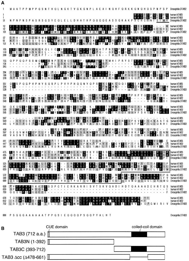
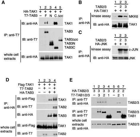
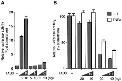
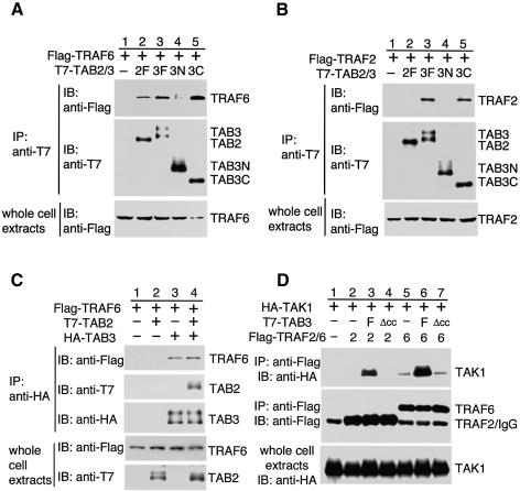
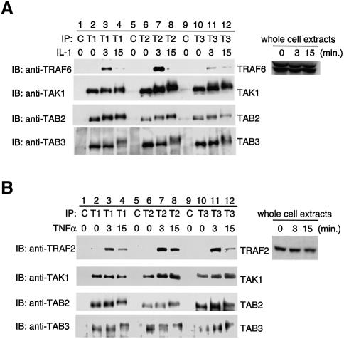
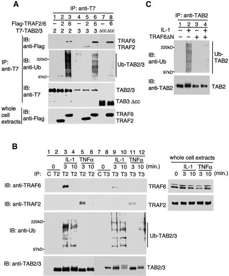
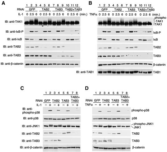
The cytokines IL-1 and TNF induce expression of a series of genes that regulate inflammation through activation of NF-kappaB signal transduction pathways. TAK1, a MAPKKK, is critical for both IL-1- and TNF-induced activation of the NF-kappaB pathway. TAB2, a TAK1-binding protein, is involved in IL-1-induced NF-kappaB activation by physically linking TAK1 to TRAF6. However, IL-1-induced activation of NF-kappaB is not impaired in TAB2-deficient embryonic fibroblasts. Here we report the identification and characterization of a novel protein designated TAB3, a TAB2-like molecule that associates with TAK1 and can activate NF-kappaB similar to TAB2. Endogenous TAB3 interacts with TRAF6 and TRAF2 in an IL-1- and a TNF-dependent manner, respectively. Further more, IL-1 signaling leads to the ubiquitination of TAB2 and TAB3 through TRAF6. Cotransfection of siRNAs directed against both TAB2 and TAB3 inhibit both IL-1- and TNF-induced activation of TAK1 and NF-kappaB. These results suggest that TAB2 and TAB3 function redundantly as mediators of TAK1 activation in IL-1 and TNF signal transduction.


Figures

None
Fig. 1. Structure of TAB3. (A) Comparison of amino acid sequences among hTAB3 (human), hTAB2 (human) and DTAB2 (Drosophila). They share the CUE domain (bold underline) and coiled-coil structure (box). Identical and conserved amino acids are indicated by black and gray boxes, respectively. DDBJ/EMBL/GenBank accession No. for hTAB3 is AY437560. (B) Schematic representation of various TAB3 constructs. Gray and black boxes indicate the CUE domain and coiled-coil structure, respectively.
None
Fig. 2. TAB3 interacts with and activates TAK1. (A) Interaction of TAB3 with TAK1. The 293 cells were transfected with plasmids encoding T7-TAB3 full-length (F), T7-TAB3N (N), T7-TAB3C (C), T7-TAB3Δcc (Δcc) and HA-TAK1 as indicated. Complexes immunoprecipitated with anti-T7 antibody were immunoblotted with anti-HA or anti-T7 antibodies. Whole-cell extracts were immunoblotted with anti-HA antibody. (B and C) Activation of TAK1 and JNK by TAB3. The 293 cells were transfected with plasmids encoding HA-TAK1, HA-JNK, T7-TAB2 (2) and T7-TAB3 (3) as indicated. HA-TAK1 or HA-JNK was immunoprecipitated with anti-HA antibody. The immunoprecipitates were subjected to an in vitro phosphorylation assay using bacterially expressed MKK6 (B) or GST-c-Jun (C) as an exogenous substrate. The immunoprecipitates were analyzed by immunoblotting with anti-HA antibody. (D) Effect of TAB2 on the interaction between TAK1 and TAB3. The 293 cells were transfected with plasmids encoding HA-TAB3, T7-TAB2 and Flag–TAK1 as indicated. Complexes immunoprecipitated with anti-HA antibody were immunoblotted with anti-Flag, anti-T7 or anti-HA antibodies. Whole-cell extracts were immunoblotted with anti-Flag or anti-T7 antibodies. (E) Interaction among TAK1-binding proteins. The 293 cells were transfected with plasmids encoding HA-TAB2 (2), HA-TAB3 (3), T7-TAB1 (1), T7-TAB2 (2) and T7-TAB3 (3) as indicated. Complexes immunoprecipitated with anti-T7 antibody were immunoblotted with anti-HA or anti-T7 antibodies. Whole-cell extracts were immunoblotted with anti-HA antibody.
None
Fig. 3. TAB3 is involved in NF-κB activation pathway. (A) Effects of TAB3 on NF-κB-dependent reporter gene activity. The 293 cells were transfected with luciferase reporter plasmid (0.1 µg) and the indicated amounts of plasmids encoding TAB3 full-length (F), TAB3N (N) and TAB3C (C). After 24 h incubation, cells were harvested and luciferase activity measured. The values shown are the average of one representative experiment in which each transfection was performed in duplicate. (B) Effects of TAB3 on IL-1- and TNFα-induced NF-κB-activation. 293IL-1RI cells were transfected with luciferase reporter plasmid (0.1 µg) and the indicated amounts of plasmids encoding TAB3N (N) and TAB3C (C). IL-1β (5 ng/ml) or TNFα (10 ng/ml) was added to each plate 3 h after transfection. Cells were harvested 24 h after transfection and luciferase activity was measured. The values shown are the average of one representative experiment in which each transfection was performed in duplicate.
None
Fig. 4. TAB3 mediates the interaction of TAK1 with TRAF6 and TRAF2. (A and B) Interaction of TAB3 with TRAF6 and TRAF2. The 293 cells were transfected with plasmids encoding T7-TAB2 full-length (2F), T7-TAB3 full-length (3F), T7-TAB3N (3N), T7-TAB3C (3C), Flag-TRAF6 and Flag-TRAF2 as indicated. Complexes immunoprecipitated with anti-T7 antibody were immunoblotted with anti-Flag or anti-T7 antibodies. Whole-cell extracts were immunoblotted with anti-Flag antibody. (C) Effect of TAB2 on the interaction between TRAF6 and TAB3. The 293 cells were transfected with plasmids encoding HA-TAB3, T7-TAB2 and Flag-TRAF6 as indicated. Complexes immunoprecipitated with anti-HA antibody were immunoblotted with anti-Flag, anti-T7 or anti-HA antibodies. Whole-cell extracts were immunoblotted with anti-Flag or anti-T7 antibodies. (D) Effect of TAB3 on the interaction of TAK1 with TRAF2 and TRAF6. The 293 cells were transfected with plasmids encoding HA-TAK1, T7-TAB3 full-length (F), T7-TAB3Δcc (Δcc), Flag-TRAF2 (2) and Flag-TRAF6 (6) as indicated. Complexes immunoprecipitated with anti-Flag antibody were immunoblotted with anti-HA or anti-Flag antibodies. Whole-cell extracts were immunoblotted with anti-HA antibody.
None
Fig. 5. Ligand-dependent association of TRAF6 and TRAF2 with TAK1, TAB2 and TAB3. 293IL-1RI cells were treated with (A) IL-1 (10 ng/ml) or (B) TNFα (10 ng/ml) for the indicated time periods. Endogenous TAK1, TAB2 and TAB3 were immunoprecipitated with anti-TAK1 (T1), anti-TAB2 (T2) and anti-TAB3 (T3) antibodies, respectively. Complexes immunoprecipitated with control IgG (C) or each antibody was immunoblotted with anti-TRAF6, anti-TRAF2, anti-TAK1, anti-TAB2 or anti-TAB3 antibodies. Whole-cell extracts were immunoblotted with anti-TRAF6 or anti-TRAF2 antibodies.
None
Fig. 6. Ubiquitination of TAB2 and TAB3. (A) Effect of TRAF2 and TRAF6 on ubiquitination of TAB2 and TAB3. 293IL-1RI cells were transfected with plasmids encoding T7-TAB2 (2), T7-TAB3 (3), T7-TAB3Δcc (Δcc), Flag-TRAF2 (2) and Flag-TRAF6 (6) as indicated. Complexes immunoprecipitated with anti-T7 antibody were immunoblotted with anti-Flag, anti-ubiquitin (anti-Ub) or anti-T7 antibodies. Whole-cell extracts were immunoblotted with anti-Flag antibody. (B) Effect of IL-1 and TNFα stimulation on ubiquitination of TAB2 and TAB3. 293IL-1RI cells were treated with IL-1 (10 ng/ml) or TNFα (20 ng/ml) for the indicated time periods. Cell extracts were subjected to immunoprecipitation with control IgG (C), anti-TAB2 (T2) or anti-TAB3 (T3) antibodies. Immunoprecipitated complexes were immunoblotted with anti-TRAF6, anti-TRAF2, anti-Ub, anti-TAB2 or anti-TAB3 antibodies. Whole-cell extracts were immunoblotted with anti-TRAF6 or anti-TRAF2 antibodies. (C) Effect of TRAF6ΔN on IL-1-induced ubiquitination of TAB2. 293IL-1RI cells were transfected with a plasmid encoding TRAF6ΔN. At 48 h post-transfection, cells were treated with IL-1 (10 ng/ml) for 10 min. Cell extracts were subjected to the immunoprecipitation with anti-TAB2 antibody. Immunoprecipitated complexes were immunoblotted with anti-Ub or anti-TAB2 antibodies.
None
Fig. 7. Effects of TAB2 and TAB3 siRNAs on IL-1 and TNF signaling pathways. HeLa cells were transfected with annealed sense and antisense 21-mer siRNA oligonucleotides directed against Jellyfish GFP (GFP; as a control siRNA), TAB2 or TAB3 using Oligofectamine: 400 nM GFP siRNA; 200 nM TAB2 siRNA + 200 nM GFP siRNA; 200 nM TAB3 siRNA + 200 nM GFP siRNA; 200 nM TAB2 siRNA + 200 nM TAB3 siRNA. At 72 h post-transfection, the cells were treated with IL-1 (10 ng/ml) (A and C) or TNFα (10 ng/ml) (B and D) for the indicated times (A and B) or for 10 min (C and D). Western blot analysis was performed on extracts prepared from these cells using antibodies directed against TAK1, phospho-IκBα (IκB-P), IκBα, TAB2, TAB3, TAB1, phospho-p38, p38 or JNK1. Western blot analysis for β-catenin indicates that an equivalent amount of protein was present in each lane.

References

    1. Baud V. and Karin,M. (2001) Signal transduction by tumor necrosis factor and its relatives. Trends Cell Biol., 11, 372–377. - PubMed
    1. Cao Z., Xiong,J., Takeuchi,M., Kurama,T. and Goeddel,D.V. (1996) TRAF6 is a signal transducer for interleukin-1. Nature, 383, 443–446. - PubMed
    1. Deng L., Wang,C., Spencer,E., Yang,L., Braun,A., You,J., Slaughter,C., Pickart,C. and Chen,Z.J. (2000) Activation of the IκB kinase complex by TRAF6 requires a dimeric ubiquitin-conjugating enzyme complex and a unique polyubiquitin chain. Cell, 103, 351–361. - PubMed
    1. Dinarello C.A. (1996) Biologic basis for interleukin-1 in disease. Blood, 87, 2095–2147. - PubMed
    1. Ghosh S. and Karin,M. (2002) Missing pieces in the NF-κB puzzle. Cell, 109, S81–S96. - PubMed

Publication types

MeSH terms