. 2003 Dec 1;22(23):6289-98.
doi: 10.1093/emboj/cdg613.

E2F7, a novel E2F featuring DP-independent repression of a subset of E2F-regulated genes

Affiliations

E2F7, a novel E2F featuring DP-independent repression of a subset of E2F-regulated genes

Luisa Di Stefano et al. EMBO J. .

Abstract

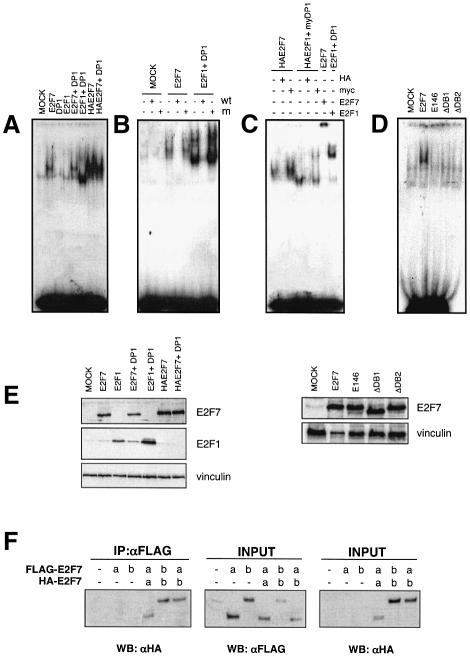
The E2F family of transcription factors play an essential role in the regulation of cell cycle progression. In a screen for E2F-regulated genes we identified a novel E2F family member, E2F7. Like the recently identified E2F-like proteins of Arabidopsis, E2F7 has two DNA binding domains and binds to the E2F DNA binding consensus site independently of DP co-factors. Consistent with being an E2F target gene, we found that the expression of E2F7 is cell cycle regulated. Ectopic expression of E2F7 results in suppression of E2F target genes and accumulation of cells in G1. Furthermore, E2F7 associates with E2F-regulated promoters in vivo, and this association increases in S phase. Interestingly, however, E2F7 binds only a subset of E2F-dependent promoters in vivo, and in agreement with this, inhibition of E2F7 expression results in specific derepression of these promoters. Taken together, these data demonstrate that E2F7 is a unique repressor of a subset of E2F target genes whose products are required for cell cycle progression.


Figures

None
Fig. 1. E2F7 contains two domains similar to the DNA binding domain of E2F proteins. (A) Intron–exon structure of E2F7a and E2F7b. (B) Domain organization of E2F7a and E2F7b compared with the other members of the E2F family. Number of amino acids is indicated on the right. Shaded boxes indicate homologous regions. cA, Cyclin A binding site; DB, DNA binding domain; DIM, dimerization domain; TA, transactivation domain; PB, pocket protein (pRB family) binding domain. (C) Amino acid sequence alignment of the DB1 and DB2 domains of E2F7 with the DNA binding domains of the other E2Fs and DPs. Grey highlighted characters indicate amino acids conserved among these proteins. (D) Expression of E2F7a and E2F7b. Western blot analysis of cell lysates derived from U2OS transfected with either pCDNA E2F7a (a), E2F7b (b) or pCDNA vector alone (–). Endogenous and ectopic E2F7 protein expression was detected by immunoblotting with an antibody to E2F7. Cell lysate (40 µg) was loaded to detect the endogenous proteins (–), whereas only 10 µg was loaded of the cell lysates prepared from cells overexpressing E2F7a and E2F7b.
None
Fig. 2. E2F7 is an E2F target gene. (A) Real-time qPCR analysis of mRNA isolated from WI38 cells expressing ER–E2F1. Cells were incubated with OHT, cycloheximide (CHX) or both for 4 h. (B) E2F1 and E2F4 associate with the E2F7 promoter in vivo. Asynchronously growing U2OS cells were treated with formaldehyde and enrichment of the E2F7 promoter sequences was tested by ChIP using the indicated antibodies. β-actin and E2F1 promoters were used respectively as negative and positive controls. The percentage of the bound promoter/total promoter present in the cells is indicated. (C) WI38 cells were serum-starved and re-stimulated to enter the cell cycle by serum addition. qPCR analysis was performed at subsequent time points to determine the E2F7 expression profile. Propidium iodide (PI) FACS analysis is shown at the top of the panel. The known E2F target gene, CCNE1, is shown as control. (D) The expression of E2F7a and E2F7b is cell growth regulated. Western blot analysis of cell lysates prepared from WI38 cells starved and released as described in panel C. Antibodies specific for E2F7 and Cyclin A2 were used as indicated. Vinculin levels were analysed to check equal loading of the gel. PI/FACS analysis is presented at the top of the panel. (E) E2F7b is cell cycle regulated. U2OS cells were synchronized in metaphase and released into the cell cycle by plating in nocodazole-free medium. Cells were harvested at the indicated times after release and analysed as in (D).
None
Fig. 3. Specific binding of E2F7 to E2F-binding sites independently of DP. (A) E2F7 binds E2F sites. U2OS cell extracts prepared from cells transfected with the indicated expression plasmids were incubated with E2F oligonucleotide and assayed for binding. (B) E2F7 binding requires the E2F consensus site. A 100-fold excess of either wild-type (w.t.) or mutant (m) cold competitor probe was added as indicated. (C) Antibody super-shifts were performed using anti-E2F1 (KH20), anti-HA (Babco), anti-Myc (9E10) and anti-E2F7 as indicated. (D) DNA binding requires intact DB1 and DB2 domains: mutant versions of E2F7 protein were assayed for binding. (E) Expression levels of the transfected constructs in U2OS cells were determined by western blotting using anti-E2F7 and anti-E2F1 (KH20) antibodies. Vinculin was used as a loading control. (F) E2F7 can homodimerize. U2OS cells were transfected with either Flag-tagged E2F7a or E2F7b alone or in combination with HA-tagged E2F7a or E2F7b. Anti-Flag antibody was used to immunoprecipitate the Flag-tagged E2Fs. Bound E2F7a and E2F7b proteins were detected by western blot using an anti-HA specific antibody. The levels of E2F7a and E2F7b expressed in transfected cells was measured by western blotting of aliquots taken before the immunoprecipitation (INPUT) using anti-Flag or anti-HA antibodies.
None
Fig. 4. E2F7 represses E2F responsive promoters. (A) Transfection assay with 6× E2F Luc reporter and increasing amounts (10, 30 and 100 ng) of E2F7a expression plasmid and (B) E2F7b expression plasmid. (C) E2F7a and E2F7b can repress transcription of the E2F1 promoter [pGL3 E2F1 (-242)].The assay was performed using 100 ng of E2F7a and E2F7b expression plasmids. (D) Repression is dependent on intact E2F sites. Transfection assay with mutant [pGL3E2F1(-242, -E2F)] E2F1constructs. (E) The DNA binding domains of E2F7 are required for repression. The effect of expressing wild-type or DNA binding mutant E2F7 proteins were evaluated on the 6× E2F luciferase promoter; 100 ng of the indicated plasmids were used for the transfections. (F) E2F7 competes with E2F1 in regulating E2F-responsive promoters. Increasing amounts of E2F1 expression plasmid (20–100 ng) were transfected in the presence or absence of constant amounts (100 ng) of E2F7 expression plasmid. All luciferase values were corrected for transfection efficiency by normalizing with β-galactosidase activity. Experiments were performed in duplicate and results were reproduced at least three times. Error bars indicate standard deviation of the mean.
None
Fig. 5. E2F7 binds to E2F responsive promoters in vivo. (A) ChIP analysis of asynchronous U2OS cells. ChIP was performed using antibodies specific for E2F1 and E2F7. Anti-Flag was used as negative control. The resulting DNA was amplified by qPCR with primers corresponding to the genes shown. β-actin promoter was used as a negative control. (B) U2OS cells were synchronized in mitosis by treating with thymidine followed by nocodazole. Cells were stimulated to re-enter the cell cycle by plating in nocodazole-free medium, harvested at the indicated time points and examined by FACS analysis. (C) ChIP analysis of U2OS cells synchronized as described in (B) was performed using the indicated antibodies as in (A).
None
Fig. 6. Increased association of E2F7 with E2F responsive promoters as cells enter the S phase of the cell cycle. (A) WI38 diploid human fibroblast were rendered quiescent and subsequently induced to enter the cell cycle by the addition of serum-containing medium. Cell cycle profiles were determined by PI/FACS. (B) ChIP was performed using the indicated antibodies as described in Figure 5A.
None
Fig. 7. E2F7 inhibits S phase entry and cell proliferation. (A) Ectopic E2F7 expression inhibits Rat1 cells from S phase entry. Serum-starved quiescent Rat1 cells were microinjected with expression plasmids for the indicated proteins. Immediately after microinjection, medium containing serum and BrdU was added, and the number of cells in S phase was determined 16 h later. (B) E2F7’s ability to inhibit S phase entry is dependent on DNA binding. Rat1 cells were microinjected with the indicated expression plasmids, and BrdU was determined as described in (A). p16 was used as a positive control. (C) Ectopic expression of E2F7 results in accumulation of cells in G1. GFP-positive U2OS cells transfected with pCMV vectors, as indicated, were analysed by PI/FACS. (D) Ectopic expression of E2F7 impairs growth. HeLa cells were transfected with indicated pCMV vectors. Cells were plated (50 000 cells/10-cm plate) and selected in neomycin for 14 days prior to crystal violet staining.
None
Fig. 8. E2F7 represses a subset of E2F-dependent genes in vivo. (A) Specific depletion of E2F7a and E2F7b by siRNA in U2OS cells. Western blot analysis of U2OS cells transfected with siRNA oligos designed to interfere with E2F7 mRNA using antibodies specific for E2F7 and vinculin (loading control). An unrelated siRNA specific for luciferase (GL3) and mock-transfected cells were used as controls. Cell lysates were prepared 24 and 48 h after transfection with the siRNA oligos. (B) E2F7 depletion results in up-regulation of expression of E2F7-specific target genes. qPCR analysis of the expression of the indicated genes 48 h after transfection of U2OS cells with siRNA to E2F7. The expression level is normalized against GL3 transfected control. Determination was performed in triplicate and the standard deviation of the mean is indicated.

Similar articles

Cited by

References

    1. Bandara L.R., Buck,V.M., Zamanian,M., Johnston,L.H. and La Thangue,N.B. (1993) Functional synergy between DP-1 and E2F-1 in the cell cycle-regulating transcription factor DRTF1/E2F. EMBO J., 12, 4317–4324. - PMC - PubMed
    1. Cartwright P., Muller,H., Wagener,C., Holm,K. and Helin,K. (1998) E2F-6: a novel member of the E2F family is an inhibitor of E2F-dependent transcription. Oncogene, 17, 611–623. - PubMed
    1. Courjal F., Louason,G., Speiser,P., Katsaros,D., Zeillinger,R. and Theillet,C. (1996) Cyclin gene amplification and overexpression in breast and ovarian cancers: evidence for the selection of cyclin D1 in breast and cyclin E in ovarian tumors. Int. J. Cancer, 69, 247–255. - PubMed
    1. Cress W.D., Johnson,D.G. and Nevins,J.R. (1993) A genetic analysis of the E2F1 gene distinguishes regulation by Rb, p107 and adenovirus E4. Mol. Cell. Biol., 13, 6314–6325. - PMC - PubMed
    1. Frank S.R., Schroeder,M., Fernandez,P., Taubert,S. and Amati,B. (2001) Binding of c-Myc to chromatin mediates mitogen-induced acetylation of histone H4 and gene activation. Genes Dev., 15, 2069–2082. - PMC - PubMed

Publication types

MeSH terms