. 2003 Dec 9;100(25):15166-71.
doi: 10.1073/pnas.2334624100. Epub 2003 Nov 21.

TRPM5 is a transient Ca2+-activated cation channel responding to rapid changes in [Ca2+]i

Affiliations

TRPM5 is a transient Ca2+-activated cation channel responding to rapid changes in [Ca2+]i

Dirk Prawitt et al. Proc Natl Acad Sci U S A. .

Abstract

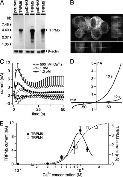
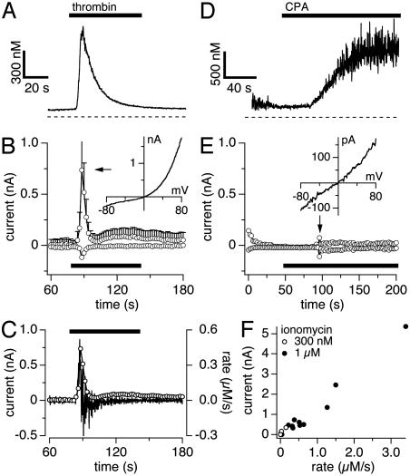
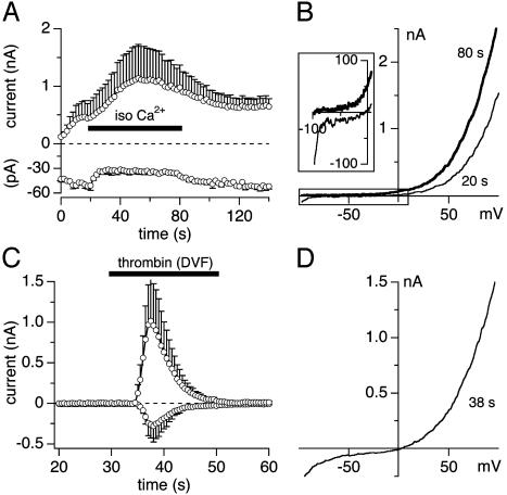
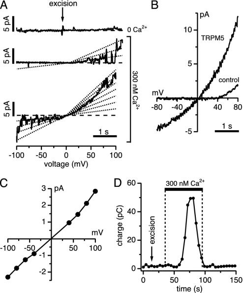
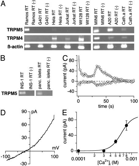
Transient receptor potential (TRP) proteins are a diverse family of proteins with structural features typical of ion channels. TRPM5, a member of the TRPM subfamily, plays an important role in taste receptors, although its activation mechanism remains controversial and its function in signal transduction is unknown. Here we characterize the functional properties of heterologously expressed human TRPM5 in HEK-293 cells. TRPM5 displays characteristics of a calcium-activated, nonselective cation channel with a unitary conductance of 25 pS. TRPM5 is a monovalent-specific, nonselective cation channel that carries Na+, K+, and Cs+ ions equally well, but not Ca2+ ions. It is directly activated by [Ca2+]i at concentrations of 0.3-1 microM, whereas higher concentrations are inhibitory, resulting in a bell-shaped dose-response curve. It activates and deactivates rapidly even during sustained elevations in [Ca2+]i, thereby inducing a transient membrane depolarization. TRPM5 does not simply mirror levels of [Ca2+]i, but instead responds to the rate of change in [Ca2+]i in that it requires rapid changes in [Ca2+]i to generate significant whole-cell currents, whereas slow elevations in [Ca2+]i to equivalent levels are ineffective. Moreover, we demonstrate that TRPM5 is not limited to taste signal transduction, because we detect the presence of TRPM5 in a variety of tissues and we identify endogenous TRPM5-like currents in a pancreatic beta cell line. TRPM5 can be activated physiologically by inositol 1,4,5-trisphosphate-producing receptor agonists, and it may therefore couple intracellular Ca2+ release to electrical activity and subsequent cellular responses.


Figures

Fig. 1.
Fig. 1.
TRPM5 is a transmembrane protein and a calcium-activated cation channel. (A) Northern blot analysis of HEK-293 cells stably transfected with pcDNA3-TRPM5 and pcDNA3. (B) Confocal laser microscopic analysis of HEK-293 cells expressing the EGFP-TRPM5 fusion protein showing significant amounts of the protein in the plasma membrane. (C) Average TRPM5 currents at -80 mV or +80 mV in HEK-293 cells perfused with intracellular solutions buffered to the indicated levels of [Ca2+]i (n = 5–20). (D) Typical I/V curve of TRPM5 currents measured 10 s or 40 s after establishment of whole-cell configuration with 500 nM [Ca2+]i. (E) Concentration–response curve of TRPM5 currents (left axis, filled circles; n = 5–20). The fit represents the product of two Boltzmann functions with an apparent EC50 for activation of 840 nM (Hill coefficient, 5). The right axis shows the dose dependence of TRPM4 currents evoked by different concentrations of intracellular [Ca2+]i (data recalculated by webmaxc, see Methods). The fit to these data yields an EC50 of 885 nM (Hill coefficient, 3.6).
Fig. 3.
Fig. 3.
TRPM5 activation depends on rate of calcium change. Data were obtained by using combined patch-clamp and fura 2 recordings. (A) Average [Ca2+]i signals in response to thrombin. (B) Average TRPM5 currents at -80 mV and +80 mV in HEK-293 cells (n = 5) evoked by thrombin (20 units/ml). (Inset) Leak-subtracted I/V curve from a typical cell. (C) Outward currents taken from B and superimposed on the differentiated calcium signal from A. (D) Typical example (n = 9) of a slow [Ca2+]i signal evoked by 50 μM cyclopiazonic acid (CPA). (E) Corresponding currents at -80 mV and +80 mV of the same cell as shown in D. The arrow points to a burst of endogenous TRPM4 activity (sample I/V in Inset). (F) TRPM5 peak currents plotted as a function of rate of change in [Ca2+]i. Data were obtained from cells stimulated by 0.3 or 1 μM ionomycin in Ca2+-free extracellular solutions. All cells included in this plot achieved at least 1 μM [Ca2+]i, albeit at different rates.
Fig. 2.
Fig. 2.
TRPM5 is a monovalent cation channel. (A) Average currents of TRPM5 at -80 mV and +80 mV (n = 3) in HEK-293 cells perfused with 500 nM [Ca2+]i and superfused with isotonic CaCl2 (120 mM). (B) Typical I/V curve before (20 s) and at the end of 80 s isotonic Ca2+ application. (Inset) Shift in reversal potential to negative values (data were leak-subtracted by using the first ramp current as template). (C) Application of divalent-free NaCl-based extracellular solution (DVF) with 1 mM Na-EDTA and 20 units/ml thrombin to activate TRPM5 (n = 5). The inactivation followed a single exponential function with a time constant τ = 5.9 s. (D) Typical I/V curve during DVF application.
Fig. 4.
Fig. 4.
Ca2+ activates TRPM5 single channels in inside-out patches. (A) TRPM5 single-channel activity in inside-out patches of HEK-293 cells. Ramps with no channel activity were used for leak correction. The top profile shows excision of the patch into 0 Ca2+ solution; the middle and bottom profiles are consecutive examples of channel activity evoked by 300 nM Ca2+.(B) Ensemble I/V curve of TRPM5 single channels (75 ramps) and control cells (WT HEK-293; 98 ramps) during 300 nM Ca2+ exposure. (C) Current–voltage relationship of TRPM5 single channels. Each point reflects the average of 15–25 events per voltage from five patches. The slope yields a channel conductance of 25 pS. (D) Average charge measured before, during, and after exposure of TRPM5-expressing patches to 300 nM Ca2+ (same patches as in AC, n = 5), assessed by integrating ramp currents between 0 mV and +100 mV.
Fig. 5.
Fig. 5.
Rat insulinoma beta cells express endogenous TRPM5-like currents. (A) Total RNA from different human and murine cell lines was isolated as described and transcribed into cDNA. RT-PCR was performed with species-specific primers for the TRPM4/Trpm4 and the TRPM5/Trpm5 genes. (B) Endogenous Trpm5 expression in pancreatic beta cells (INS-1) and in whole human pancreatic islets assessed as in A.(C) Whole-cell currents at -80 mV and +80 mV in a typical INS-1 cell (n = 8) perfused with 800 nM [Ca2+]i. The delay of TRPM5 activation varied between cells; the average delay at 500 nM [Ca2+]i = 63 ± 11s(n = 3) and at 800 nM [Ca2+]i = 53 ± 10s(n = 8). The initial drop of currents seen after break-in is due to the inactivation of K+. Data were leak-corrected by using the 12th ramp current as template. (D) Current–voltage relationship of TRPM5-like currents (n = 8). Data were leak-corrected by subtracting an appropriate control ramp before current activation. (E) Dose–response curve of endogenous TRPM5-like currents in INS-1 cells (n = 3–8); 1 μM [Ca2+]i caused large additional Ca2+-activated currents (n = 6; data not shown), which prevented accurate assessment of TRPM5 in isolation.

References

    1. Harteneck, C., Plant, T. D. & Schultz, G. (2000) Trends Neurosci. 23, 159-166. - PubMed
    1. Clapham, D. E., Runnels, L. W. & Strubing, C. (2001) Nat. Rev. Neurosci. 2, 387-396. - PubMed
    1. Minke, B. & Cook, B. (2002) Physiol. Rev. 82, 429-472. - PubMed
    1. Montell, C., Birnbaumer, L. & Flockerzi, V. (2002) Cell 108, 595-598. - PubMed
    1. Montell, C., Birnbaumer, L., Flockerzi, V., Bindels, R. J., Bruford, E. A., Caterina, M. J., Clapham, D. E., Harteneck, C., Heller, S., Julius, D., et al. (2002) Mol. Cell 9, 229-231. - PubMed

Publication types

MeSH terms