. 2003 Dec 9;100(25):14926-31.
doi: 10.1073/pnas.2434383100. Epub 2003 Nov 21.

Hematopoietic progenitors express neural genes

Affiliations

Hematopoietic progenitors express neural genes

James Goolsby et al. Proc Natl Acad Sci U S A. .

Abstract

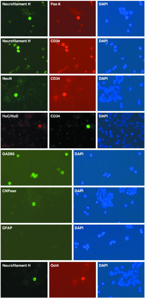
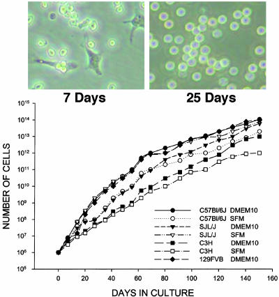
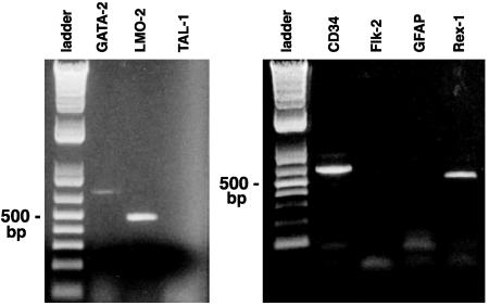
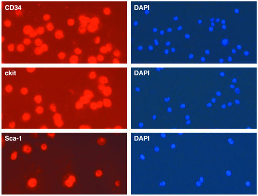
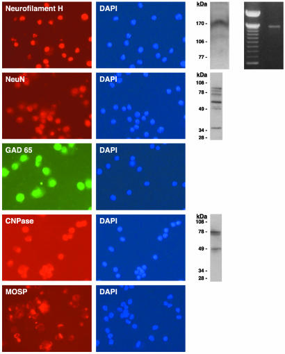
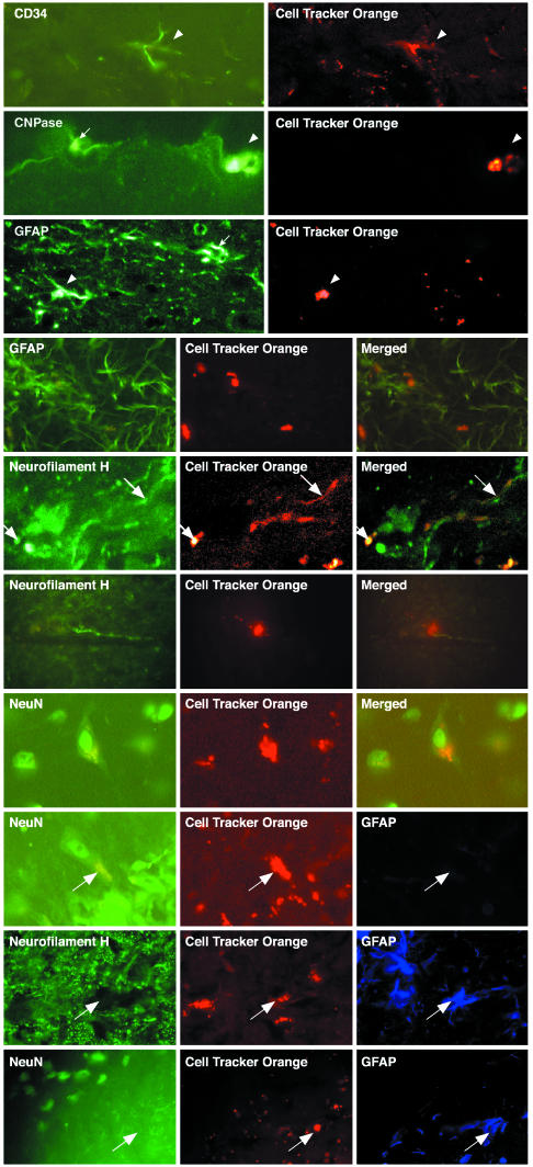
Bone marrow, or cells selected from bone marrow, were reported recently to give rise to cells with a neural phenotype after in vitro treatment with neural-inducing factors or after delivery into the brain. However, we showed previously that untreated bone marrow cells express products of the neural myelin basic protein gene, and we demonstrate here that a subset of ex vivo bone marrow cells expresses the neurogenic transcription factor Pax-6 as well as neuronal genes encoding neurofilament H, NeuN (neuronal nuclear protein), HuC/HuD (Hu-antigen C/Hu-antigen D), and GAD65 (glutamic acid decarboxylase 65), as well as the oligodendroglial gene encoding CNPase (2',3' cyclic nucleotide 3'-phosphohydrolase). In contrast, astroglial glial fibrillary acidic protein (GFAP) was not detected. These cells also were CD34+, a marker of hematopoietic stem cells. Cultures of these highly proliferative CD34+ cells, derived from adult mouse bone marrow, uniformly displayed a phenotype comparable with that of hematopoietic progenitor cells (CD45+, CD34+, Sca-1+, AA4.1+, cKit+, GATA-2+, and LMO-2+). The neuronal and oligodendroglial genes expressed in ex vivo bone marrow also were expressed in all cultured CD34+ cells, and GFAP was not observed. After CD34+ cell transplantation into adult brain, neuronal or oligodendroglial markers segregated into distinct nonoverlapping cell populations, whereas astroglial GFAP appeared, in the absence of other neural markers, in a separate set of implanted cells. Thus, neuronal and oligodendroglial gene products are present in a subset of bone marrow cells, and the expression of these genes can be regulated in brain. The fact that these CD34+ cells also express transcription factors (Rex-1 and Oct-4) that are found in early development elicits the hypothesis that they may be pluripotent embryonic-like stem cells.


Figures

Fig. 1.
Fig. 1.
Immunocytochemical detection of neural gene expression in a subset of adult mouse whole ex vivo bone marrow (see Materials and Methods). Double immunocytochemical detection of neurofilament H and Pax-6 was performed in the same subset of bone marrow cells, and expression of neuronal neurofilament H, NeuN, and HuC/HuD was present in a subset of CD34+ bone marrow cells. GAD65, an enzyme responsible for synthesis of a major neurotransmitter, also was present in a subset of bone marrow cells. Oligodendroglial CNPase was present in a subset of bone marrow cells, whereas the astroglial marker GFAP was not detected on ex vivo bone marrow. Neurofilament H and Oct-4 were found in the same subset of ex vivo bone marrow cells. DAPI stains of the nuclei of all cells are shown.
Fig. 2.
Fig. 2.
Long-term cultures of CD34+ cells from adult mouse bone marrow. (Upper) Photomicrographs of bone marrow cells at days 7 and 25. (Lower) Growth curves of cells from adult bone marrow of C57BL/6J, C3H, SJL/J, and FVB-129 mice in serum-containing and serum-free media. Growth curves of cells from the four strains of mice in serum-containing and serum-free media were comparable.
Fig. 4.
Fig. 4.
Detection of RT-PCR products of mRNA from a pure population of CD34+ cells derived from adult C57BL/6J bone marrow after 6 weeks in culture. The first lane of each panel shows a 500-bp DNA ladder. (Left) GATA-2 and LMO-2 are hematopoietic stem cell transcription factors. TAL-1 is a lymphocyte transcription factor. (Right) CD34 is a marker of hematopoietic stem cells. FLK-2 is a tyrosine kinase receptor present in hematopoietic stem cells with short-term multilineage reconstitution potential but not long-term potential. GFAP is a marker of differentiated astroglial cells. Rex-1 is a transcription factor in early embryonic stem cells. Equal quantities of RNA were used for each lane from a common pool of CD34+ cells in culture. CD34 was used as a positive control.
Fig. 3.
Fig. 3.
Immunocytochemical detection of CD34, cKit, and Sca-1 on all cells in 6-week cultures of adult C57BL/6J bone marrow. All cells in CD34+ cultures also expressed cKit and Sca-1.
Fig. 5.
Fig. 5.
Neural gene expression in adult mouse bone marrow cells cultured for 6–10 weeks. The three neuronal genes were detected as follows: neurofilament H was detected by immunocytochemistry, Western blot analysis, and RT-PCR; NeuN was detected by immunocytochemistry and Western blot; and GAD65 was detected by immunocytochemistry. The two oligodendroglial genes were detected as follows: CNPase was detected by immunocytochemistry and Western blot, and MOSP was detected by immunocytochemistry. Except for MOSP, which is a cell-surface molecule expressed in caps or rings in the plasma membrane, all markers are located inside the cells. DAPI stains of the nuclei of all cells are shown.
Fig. 6.
Fig. 6.
Immunohistochemical analysis by laser confocal microscopy of gene expression by CTO-labeled cultured CD34+, Sca-1+, AA4.1+, and cKit+ adult C57BL/6J mouse bone marrow cells transplanted into adult C57BL/6J mouse brain hippocampus and striatum. First row, CTO-labeled cells continue to express CD34 6 weeks after implantation into adult brain, and host brain cells fail to express CD34. Second row, oligodendroglial CNPase (transplanted cell, arrowhead; host cell, arrow). Third and fourth rows, astroglial GFAP (transplanted cell, arrowhead; host cell, arrow). Fifth and sixth rows, neuronal neurofilament H. Seventh row, NeuN expression in CTO-labeled adult mouse bone marrow cells 1 year after implantation into adult mouse brain. The last three rows show that transplanted CTO-labeled cells express either astroglial (GFAP) or neuronal (neurofilament H and NeuN) proteins but not both; arrows show the same cell in each row triple-labeled 1 year after implantation.

References

    1. Brazelton, T. R., Rossi, F. M., Keshet, G. I. & Blau, H. M. (2000) Science 290, 1775–1779. - PubMed
    1. Mezey, E., Chandross, K. J., Harta, G., Maki, R. A. & McKercher, S. R. (2000) Science 290, 1779–1782. - PubMed
    1. Makar, T. K., Wilt, S., Dong, Z., Fishman, P., Mouradian, M. M. & Dhib-Jalbut, S. (2002) J. Interferon Cytokine Res. 22, 783–791. - PubMed
    1. Hess, D. C., Hill, W. D., Martin-Studdard, A., Carroll, J., Brailer, J. & Carothers, J. (2002) Stroke (Dallas) 33, 1362–1368. - PubMed
    1. Bonilla, S., Alaroon, P., Villaverdi, R. Aparicio, P., Silva, A. & Martinez, S. (2002) Eur. J. Neurosci. 15, 575–582. - PubMed

Publication types

MeSH terms

Substances