. 2003 Dec 9;100(25):15065-70.
doi: 10.1073/pnas.2436329100. Epub 2003 Nov 21.

P53 hot-spot mutants are resistant to ubiquitin-independent degradation by increased binding to NAD(P)H:quinone oxidoreductase 1

Affiliations

P53 hot-spot mutants are resistant to ubiquitin-independent degradation by increased binding to NAD(P)H:quinone oxidoreductase 1

Gad Asher et al. Proc Natl Acad Sci U S A. .

Abstract

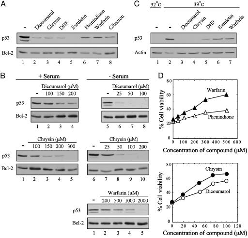
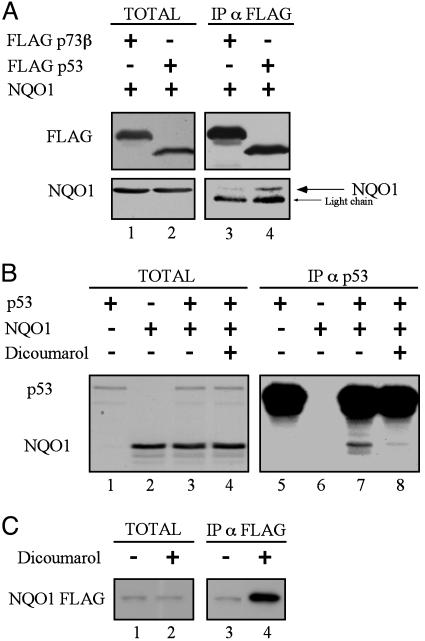
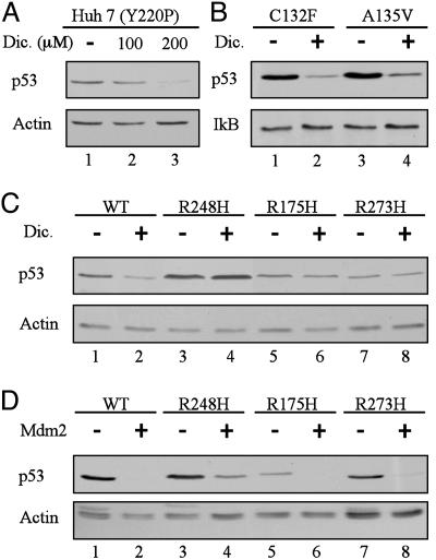
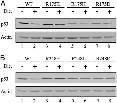
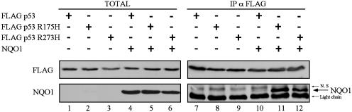
Proteasomal degradation of p53 is mediated by two alternative pathways that are either dependent or independent of both Mdm2 and ubiquitin. The ubiquitin-independent pathway is regulated by NAD(P)H: quinone oxidoreductase 1 (NQO1) that stabilizes p53. The NQO1 inhibitor dicoumarol induces ubiquitin-independent p53 degradation. We now show that, like dicoumarol, several other coumarin and flavone inhibitors of NQO1 activity, which compete with NAD(P)H for binding to NQO1, induced ubiquitin-independent p53 degradation and inhibited wild-type p53-mediated apoptosis. Although wild-type p53 and several p53 mutants were sensitive to dicoumarol-induced degradation, the most frequent "hot-spot" p53 mutants in human cancer, R175H, R248H, and R273H, were resistant to dicoumarol-induced degradation, but remained sensitive to Mdm2-ubiquitin-mediated degradation. The two alternative pathways for p53 degradation thus have different p53 structural requirements. Further mutational analysis showed that arginines at positions 175 and 248 were essential for dicoumarol-induced p53 degradation. NQO1 bound to wild-type p53 and dicoumarol, which induced a conformational change in NQO1, inhibited this binding. Compared with wild-type p53, the hot-spot p53 mutants showed increased binding to NQO1, which can explain their resistance to dicoumarol-induced degradation. NQO1 thus has an important role in stabilizing hot-spot p53 mutant proteins in human cancer.


Figures

Fig. 1.
Fig. 1.
Degradation of p53 and inhibition of p53-dependent apoptosis with different NQO1 inhibitors. (A) M1-t-p53 cells were cultured in DMEM plus serum for 5 h without (-) or with 300 μM dicoumarol, chrysin, or DHF; 2 mM phenindione, esculetin, or warfarin; or 1 mM cibacron blue. (B) M1-t-p53 cells were cultured for 5 h in DMEM with (+) or without (-) serum and without (-) or with different concentrations of dicoumarol, warfarin, or chrysin. (C) A31N-ts20 cells were preincubated at 32°C (lane 1) or 39°C for 24 h (lanes 2-7). Cells (lanes 2-7) were then incubated at 39°C for additional 5 h without (-) or with 200 μM dicoumarol or chrysin; 500 μM DHF; and 2 mM esculetin or warfarin. Immunoblot analysis was carried out by using mouse monoclonal anti-p53 antibody (Pab 240 in A and B or Pab 248 plus Pab 421 in C), hamster anti-Bcl-2, or mouse monoclonal anti-actin. (D) M1-t-p53 cells were cultured at 32°C without or with different concentrations of chrysin, dicoumarol, phenindione, or warfarin, and cell viability was determined after 23 h.
Fig. 2.
Fig. 2.
Binding of wild-type p53 to NQO1 is inhibited by dicoumarol. (A) 293 human kidney cells were transiently transfected with pEFIRES FLAG p73β or pRc/CMV FLAG p53 with pEFIRES wild-type NQO1. Extracts were electrophoresed either before immunoprecipitation (TOTAL) or after immunoprecipitation of FLAG p73β or FLAG p53 with anti-FLAG agarose beads (IP α FLAG). Immunoblot analysis was carried out with mouse monoclonal anti-FLAG antibody, and the blots were then stripped and reprobed with goat anti-NQO1 antibody. (B) In vitro reticulocyte lysate translated [35S]methionine-labeled wild-type p53 and NQO1 were incubated alone or mixed together without (-) or with (+) 300 μM dicoumarol (TOTAL). p53 was immunoprecipitated (IP α p53) with mouse monoclonal Pab 1801 anti-human p53 and NQO1 and p53 were detected by autoradiography. (C) In vitro reticulocyte lysate-translated, [35S]methionine-labeled NQO1 FLAG was incubated for 1 h in Nonidet P-40 buffer without (-) or with (+) 300 μM dicoumarol (TOTAL). NQO1 FLAG was immunoprecipitated with anti-FLAG agarose beads (IP α FLAG), and NQO1 was detected by autoradiography.
Fig. 3.
Fig. 3.
p53 mutants R248H, R175H, and R273H are resistant to dicoumarol-induced degradation but sensitive to Mdm2-mediated degradation. (A) Huh 7 cells that carry the Y220P mutant p53 were cultured for 5 h without (-) or with 100 or 200 μM dicoumarol (Dic.). (B) M1 myeloid leukemic cells that carry the C132F or A135V mouse mutant p53 cells were incubated without (-) or with (+) 300 μM dicoumarol for 5 h. (C) HCT116 p53 null cells were transiently transfected with pRc/CMV human wild-type p53 or the p53 mutants R248H, R175H, or R273H. Twenty-four hours after transfection, cells were cultured for 5 h without (-) or with (+) 300 μM dicoumarol. (D) HCT116 p53 null cells were transiently transfected with pRc/CMV human wild-type p53 or the p53 mutants R248H, R175H, or R273H without (-) or with (+) pCOC-mouse mdm2 × 2. Protein extraction and immunoblot analysis were carried out as described (5) by using mouse monoclonal anti-human p53 (Pab 1801) or anti-mouse and human p53 (Pab 240) antibody. The blots were then stripped and reprobed with mouse monoclonal anti-actin or rabbit anti-IκB antibody.
Fig. 4.
Fig. 4.
Arginines at positions 175 or 248 are essential for dicoumarol-induced degradation of p53. HCT116 p53 null cells were transiently transfected with pRc/CMV human wild-type p53, or the p53 mutants R175K, R175H, or R175D (A) or with pRc/CMV human p53 mutants R248H, R248L, or R248P (B). Twenty-four hours after transfection, cells were cultured for 5 h without (-) or with (+) 300 μM dicoumarol. Immunoblot analysis was carried out by using mouse monoclonal anti-human p53 (Pab 1801) and mouse monoclonal anti-actin antibody.
Fig. 5.
Fig. 5.
p53 mutants R175H and R273H bind wild-type NQO1 in vivo with higher affinity than wild-type p53. 293 human kidney cells were transiently transfected with pRc/CMV FLAG wild-type p53 or with the p53 mutants R175H or R273H alone or cotransfected with pEFIRES NQO1. Extracts were electrophoresed either before immunoprecipitation (TOTAL) or after immunoprecipitation of FLAG p53 with anti-FLAG agarose beads (IP α FLAG). Immunoblot analysis was carried out with mouse monoclonal anti-FLAG antibody, and the blots were then stripped and reprobed with goat anti-NQO1 antibody. N.S, nonspecific band.

Similar articles

Cited by

References

    1. Vogelstein, B., Lane, D. & Levine, A. J. (2000) Nature 408, 307-310. - PubMed
    1. Haupt, Y., Maya, R., Kazaz, A. & Oren, M. (1997) Nature 387, 296-299. - PubMed
    1. Kubbutat, M. H., Jones, S. N. & Vousden, K. H. (1997) Nature 387, 299-303. - PubMed
    1. Asher, G., Lotem, J., Sachs, L., Kahana, C. & Shaul, Y. (2002) Proc. Natl. Acad. Sci. USA 99, 13125-13130. - PMC - PubMed
    1. Asher, G., Lotem, J., Cohen, B., Sachs, L. & Shaul, Y. (2001) Proc. Natl. Acad. Sci. USA 98, 1188-1193. - PMC - PubMed

Publication types

MeSH terms