. 2003 Nov;203(5):435-50.
doi: 10.1046/j.1469-7580.2003.00239.x.

Seasonal changes in the lower jaw skeleton in male Atlantic salmon (Salmo salar L.): remodelling and regression of the kype after spawning

Affiliations

Seasonal changes in the lower jaw skeleton in male Atlantic salmon (Salmo salar L.): remodelling and regression of the kype after spawning

P Eckhard Witten et al. J Anat. 2003 Nov.

Abstract

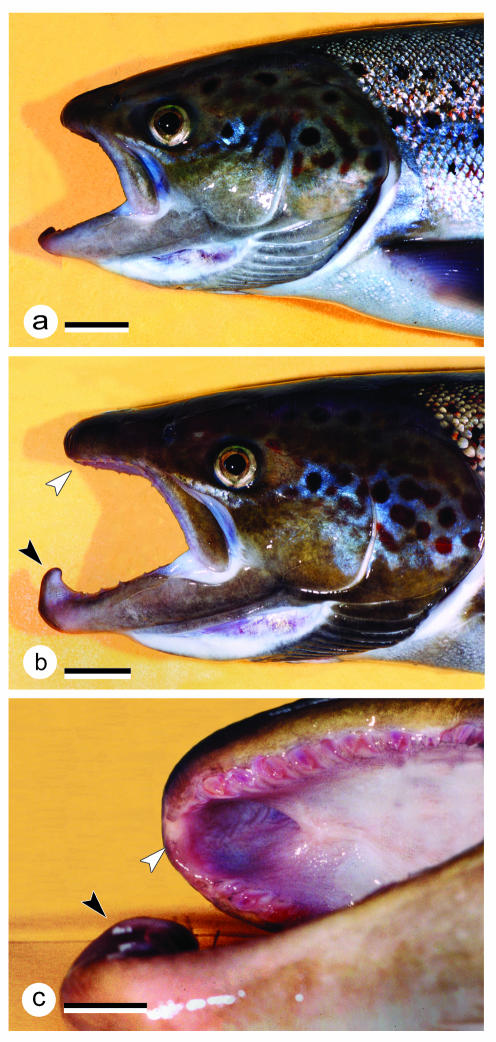
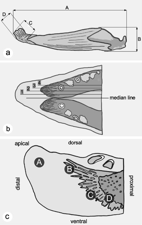
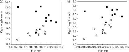
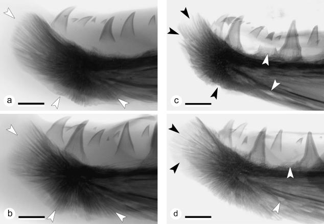
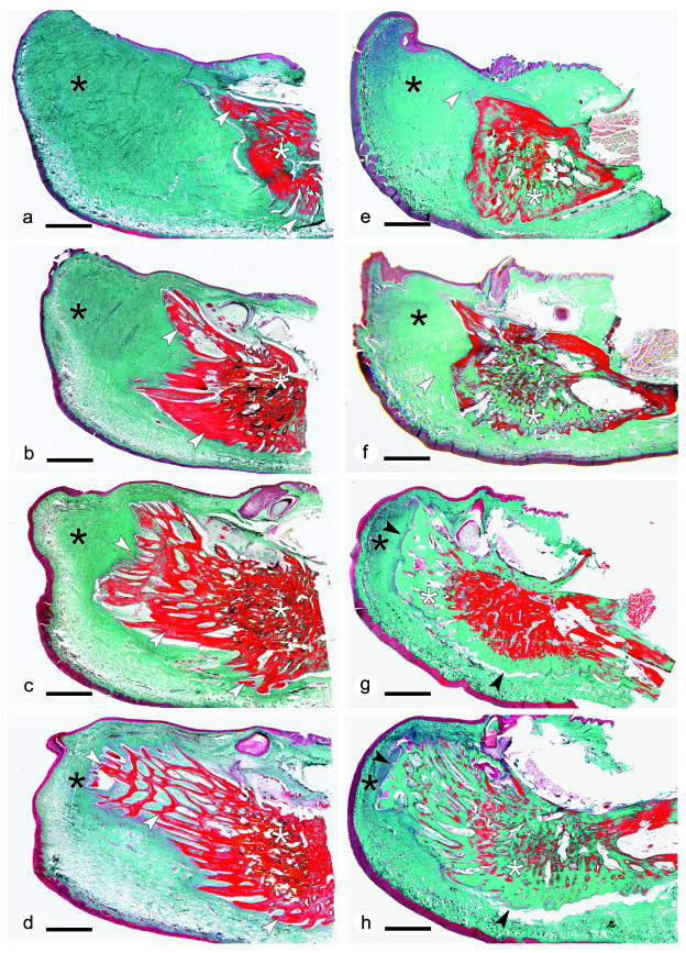
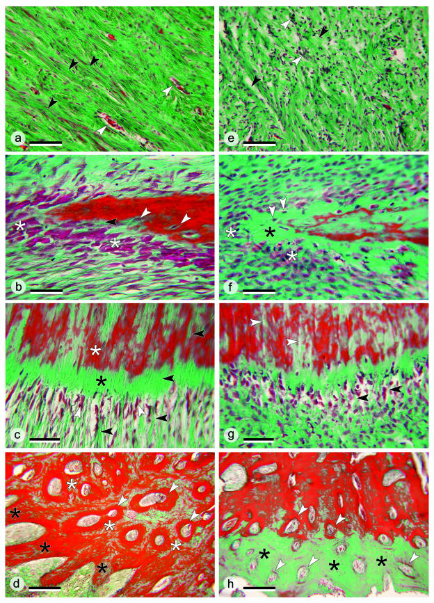
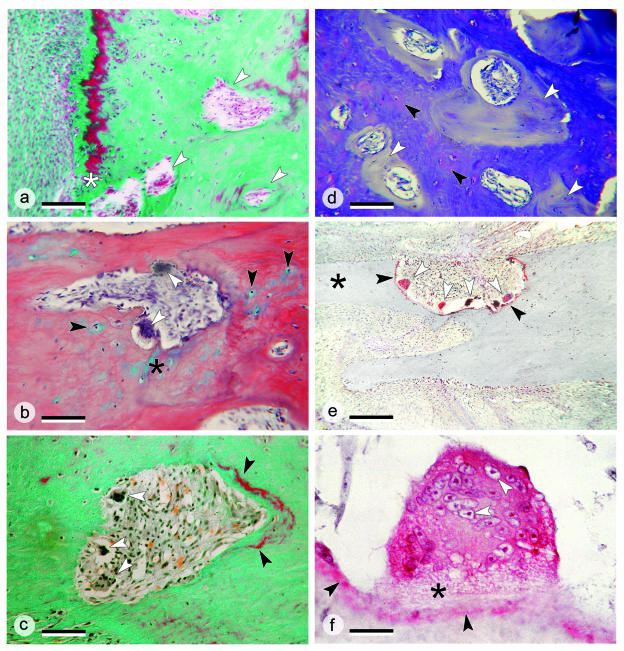
The return of Atlantic salmon (Salmon salar) to their home river for spawning coincides with drastic skeletal alterations in both sexes. Most prominent is the development of a kype (hook) at the tip of the lower jaw in males. Salmon that survive spawning have to cope with the kype throughout their life, unless it disappears after spawning, as was suggested in the early literature. To understand the fate of the kype skeleton, we compared morphological and histological features of kypes from pre-spawned mature anadromous males (grilse) with post-spawned males (kelts). The kype of male grilse is supported by fast-growing skeletal needles that differ from regular dentary bone. In kelts, growth of the kype skeleton has stopped and skeletal needles are resorbed apically by osteoclasts. Simultaneously, and despite the critical physiological condition of the animals, proximal parts of the kype skeleton are remodelled and converted into regular dentary bone. Apical resorption of the skeleton explains reports of a decrease of the kype in kelts. The conversion of basal kype skeleton into regular dentary bone contributes to the elongation of the dentary and probably also to the development of a larger kype in repetitive spawning males.


Figures

Fig. 1
Fig. 1
Heads of sexually mature female (a) and male (b) salmon grilse, ×0.6; scale bar = 20 mm. Comparison of the female with the male shows the prolongation of the jaws in males that takes place in the course of the animals’ return to their home rivers for spawning. In addition, males develop a kype (hook) at the tip of the lower jaw. The kype fits into a hole in the upper jaw, located between the two halves of the premaxilla (c). The black arrowhead indicates the kype, the white arrowhead points to the hole in the upper jaw.
Fig. 2
Fig. 2
Measurements, levels of sagittal sections and areas of the kype that have been analysed. (a) Measurements taken from the lower jaw and the kype of grilse and kelts; values are given in Table 1. A, total length of the lower jaw (dentary, angular and articular). B, maximal height of the lower jaw. C, length of the kype skeleton. D, height of the kype skeleton. (b) Levels of sagittal sections at the tip of the lower jaw in grilse and kelts. Level 1 corresponds to Fig. 5(a,e), level 2 to Fig. 5(b,f), level 3 to Fig. 5(c,g), and level 4 to Fig. 5(d,h). (c) Areas of the salmon kype in grilse and kelts compared via the histological analysis. A, the apical connective tissue of the kype, corresponding to Fig. 6(a,e). B, the growth zone of the apically directed skeletal needles, corresponding to Fig. 6(b,f). C, the growth zone of the ventral kype skeleton, corresponding to Fig. 6(c,g). D, the transitional zone between compact bone of the dentary and the chondroid bone of the kype skeleton, corresponding to Fig. 6(d,h).
Fig. 3
Fig. 3
The distribution (mm) of length (a) and height (b) of the kype skeleton in grilse (black squares) and kelts (grey circles) from all 20 animals, plotted against fork length (mm) to show variation in the dimensions of the kype among individuals and the overall reduction of the size of the kype skeleton in kelts when compared with grilse.
Fig. 4
Fig. 4
Radiographs of the lower jaw tips from two grilse (a,b) and two kelts (c,d); colours inverted, ×3.5, scale bars = 4 mm. (a) X-ray of the tip of the lower jaw of a male grilse (FL = 58.7 cm, W = 2.2 kg, K = 1.04). The kype is supported by skeletal needles that extend apically, ventrally and ventro-caudally (white arrows). As shown by this animal the kype skeleton in grilse can be long and narrow with a rounded tip. Comparison with (b) reveals the variability of the kype skeleton among individuals. (b) X-ray of the tip of the lower jaw of a male grilse (FL = 61.5 cm, W = 2.5 kg, K = 1.08). The kype supporting skeletal needles extend apically, ventrally and ventro-caudally (white arrows). This kype skeleton is broad with a flattened tip (compare with a). (c) X-ray from the tip of the lower jaw of a male kelt (FL = 57.8 cm, W = 1.3 kg, K = 0.68), showing reduction of the kype skeleton by loss of apical, ventral and ventro-caudal skeletal needles (black arrows). Translucent zones in the dentary bone and the loss of teeth (white arrows) suggest that not only the kype skeleton is being resorbed. In comparison with the kype in grilse, the connective tissue is less radio-opaque. (d) X-ray of the reduced kype skeleton of a male kelt (FL = 57.4 cm, W = 1.2 kg, K = 0.63). An area of less radio-opaque skeletal needles at the tip of the kype skeleton (black arrows) indicates demineralization of skeletal needles rather than their removal. White arrows point to a site of tooth loss (upper arrow) and to a hole in the dentary bone that is not present in grilse.
Fig. 5
Fig. 5
(a–d) Sagittal sections through the tip of the lower jaw of a male grilse (the same animal as shown in Fig. 4a), displaying connective tissue (black asterisks, green stain), the kype skeleton (made from skeletal needles, white arrows) and the compact bone of the dentary (white asterisks). Apical skeletal needles are free; proximal skeletal needles anastomose into a spongiosa-like meshwork. Apical skeletal needles are longer than ventral needles. The deep red-stained bone matrix suggests that the tissue is well mineralized. Masson's trichrome staining; ×6; scale bar = 2 mm. (e–h) Sagittal sections through the tip of the lower jaw of a male kelt (the same animal as shown in Fig. 4c), corresponding to the sections through the tip of the lower jaw in grilse shown in (a–d). The comparison with grilse reveals reduction of connective tissue (black asterisks, green stain) and the loss of apical skeletal needles (white arrows). In the absence of skeletal needles the surface of the kype skeleton is smooth (black arrows). Similar to the pattern seen in X-rays (Fig. 4c), the absence of red staining in large parts of the bone matrix (white asterisks) suggests that in comparison with grilse, the skeleton in kelts is less densely mineralized. Masson's trichrome staining; ×6; scale bar = 2 mm.
Fig. 6
Fig. 6
(a–d) Histological details in the kype of grilse; sections stained with Masson's trichrome. (a) Connective tissue supporting the kype of a grilse from area A in Fig. 2(c). Dense fibre bundles (black arrows) are arranged in parallel with blood vessels between (white arrows). ×160; scale bar = 100 µm. (b) Apical skeletal needle of the kype of a grilse from area B in Fig. 2(c). Numerous large osteoblasts (white asterisk) are located at the tip of a skeletal needle. The enclosure of osteoblasts (white arrows) into newly synthesized bone matrix and a small osteoid seam (black arrow, green stain) both indicate rapid bone formation and mineralization. ×400; scale bar = 40 µm. (c) Ventral part of the kype skeleton of a grilse from area C of Fig. 2(c). Osteoblasts (white arrows) are located between collagen fibre bundles (black arrows), adjoined by osteoid (black asterisk) and mineralized skeletal matrix (white asterisk). Continuation of collagen fibre bundles inside the bone matrix defines this part of the kype skeleton as Sharpey-fibre bone. ×250; scale bar = 60 µm. (d) The border between the kype skeleton and the compact bone of the dentary in a grilse (area D, Fig. 2c). Kype skeletal needles are composed of chondroid bone (black asterisks) adjoined by compact bone of the dentary (white asterisks) composed from osteons (white arrows). ×63; scale bar = 250 µm. (e–h) Histological details in the kype of kelts; sections stained with Masson's trichrome. (e) Apically located kype supporting connective tissue of a kelt (Fig. 2c, area A). Compared with grilse (a), collagen fibre bundles are much less organized and less dense (black arrows). Large numbers of unmasked cell nuclei are visible (white arrows). ×160; scale bar = 100 µm. (f) Apical skeletal needles of a kelt (Fig. 2c, area B). Compared with grilse (b) small and diminished numbers of osteoblasts (white asterisks) indicate that bone formation has largely stopped. The apparently ongoing transformation of osteoblasts into osteocytes (white arrows) suggests that kelts might still maintain slow bone formation, even if the matrix (black asterisk) is not mineralized. ×400; scale bar = 40 µm. (g) The ventral part of the kype skeleton of a kelt (Fig. 2c, area C). Compared with grilse (Fig. 6c) the shape of the osteoblasts has changed, similar to the changes shown in (f). Sharpey's fibres (black arrows) are less regularly arranged and appear less abundant. Gaps inside the bone matrix (white arrows) could be linked to the reduction of Sharpey's fibres. ×250; scale bar = 60 µm. (h) The border between the kype skeleton and compact bone of the dentary in a kelt (Fig. 2c, area D). Through the loss of skeletal needles and the ongoing development of osteons (white arrows), compact bone now reaches almost to the surface of the former kype skeleton. The absence of red staining in large parts of the bone matrix (black asterisks) suggests poor mineralization. ×63; scale bar = 250 µm.
Fig. 7
Fig. 7
Details of the remodelling of the kype skeleton. (a) Higher magnification of the border between the kype skeleton and compact bone of the dentary in the kelt shown in Fig. 6(h). The ventral bone surface is to the left. Osteons arise in the rudiments of the kype skeleton (white arrows), a process that starts directly beneath the bone surface (white asterisk). Masson's Trichrome staining; ×100; scale bar = 150 µm. (b) Removal of apical kype skeletal tissue in a kelt by osteoclasts (white arrows). The tissue that is removed is a chondroid bone, i.e. a bone that contains chondrocytes (black arrows) and cartilage matrix (black asterisk). HBQ staining; ×250; scale bar = 60 µm. (c) Removal of kype skeletal tissue in a kelt by osteoclasts (white arrows) and deposition of newly mineralized bone (black arrows) as part of the process of converting kype skeletal tissue into compact bone. The ventral bone surface is located at the bottom left. Masson's Trichrome staining; ×250; scale bar = 60 µm. (d) Toluidine blue staining visualizes the transformation of kype skeletal tissue (seen as metachromatic chondroid bone, black arrows) into non-metachromatic compact bone (white arrows). Toluidine blue staining; ×100; scale bar = 150 µm. (e) The base of a skeletal needle (black asterisk) from the kype of a kelt is removed by bone resorption carried out by numerous large multinucleated osteoclasts (white arrows), which express the osteoclast marker enzyme tartrate-resistant acid phosphatase (TRAP, shown in red). The enzyme is released by the osteoclasts and thus also appears on the bone surface (black arrow). TRAP staining; ×100; scale bar = 150 µm. (f) Details of a bone-resorbing giant cell showing high intracellular TRAP activity in red, numerous cell nuclei (white arrows), and the osteoclasts’ ruffled border (black asterisk). Also, TRAP appears at the bone surface (black arrows). TRAP staining; ×1000; scale bar = 15 µm.

Similar articles

Cited by

References

    1. Arndt SKA, Benfey TJ, Cunjak RA. Effect of temporary reductions in feeding on protein synthesis and energy storage of juvenile Atlantic salmon. J. Fish. Biol. 1996;49:257–276.
    1. Ashhurst DE. Macromolecular synthesis and mechanical stability during fracture repair. In: Hall BK, editor. Bone, Vol. 5: Fracture Repair and Regeneration. Boca Raton: CRC Press; 1992. pp. 61–121.
    1. Baker DM, Larsen DA, Swanson P, Dickhoff WW. Long-term peripheral treatment of immature coho salmon (Oncorhynchus kisutch) with human leptin has no clear physiologic effect. Gen. Comp. Endocrinol. 2000;118:134–138. - PubMed
    1. Benjamin M, Rufai A, Ralphs JR. The mechanism of formation of bony spurs (enthesophytes) in the Achilles tendon. Arthritis Rheum. 2000;43:576–583. - PubMed
    1. Beresford WA. Chondroid Bone, Secondary Cartilage and Metaplasia. Baltimore: Urban & Schwarzenberg; 1981.

Publication types

LinkOut - more resources