Observations on the vomeronasal organ of prenatal Tarsius bancanus borneanus with implications for ancestral morphology
- PMID: 14635801
- PMCID: PMC1571184
- DOI: 10.1046/j.1469-7580.2003.00229.x
Observations on the vomeronasal organ of prenatal Tarsius bancanus borneanus with implications for ancestral morphology
Abstract
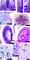
Adult primates have at least five known phenotypes of vomeronasal organ (VNO), ranging from the typical morphology seen in most other mammals to complete absence. With such morphological disparity, the phylogenetic value and any inferences on ancestral VNO morphology of the primate VNO are left uncertain. The present study investigated the VNO of embryonic and fetal Tarsius bancanus borneanus (n = 4) in comparison with prenatal specimens from four other species of primates in an effort to clarify adult morphological variations. In all except one of the fetal primates, the VNO communicated to the nasopalatine duct. One exception occurred in the largest fetal Tarsius (25 mm crown-rump length), in which the VNO communicated with the nasal cavity alone. The vomeronasal neuroepithelium was well differentiated from a thinner, non-sensory epithelium in all Tarsius and New World monkeys studied, as well as late embryonic and fetal Microcebus myoxinus. In anterior sections, this neuroepithelium was found in a more superior location in Tarsius and New World monkeys compared with Microcebus myoxinus. In all primates, masses of cell bodies were found superior to the VNO, intermingled with nerve fibres. These morphologically resembled luteinizing hormone-releasing hormone neurons described in other mammals, including humans, suggesting that a primitive association of these neurons with the VNO may exist in all primate taxa. The present study revealed that prenatal similarities exist in Tarsius and New World primates in VNO epithelial morphology. However, these are transient stages of morphology. If tarsiers and anthropoids do represent a clade (Haplorhini), then the atypical morphology seen in adult tarsiers and New World monkeys probably represents the adult VNO morphology of a haplorhine common ancestor.
Figures


Similar articles
-
Ontogeny of the nasopalatine duct in primates.Anat Rec A Discov Mol Cell Evol Biol. 2003 Sep;274(1):862-9. doi: 10.1002/ar.a.10101. Anat Rec A Discov Mol Cell Evol Biol. 2003. PMID: 12923897
-
Expression of neuron-specific markers by the vomeronasal neuroepithelium in six species of primates.Anat Rec A Discov Mol Cell Evol Biol. 2004 Nov;281(1):1190-200. doi: 10.1002/ar.a.20124. Anat Rec A Discov Mol Cell Evol Biol. 2004. PMID: 15470676
-
Histological definition of the vomeronasal organ in humans and chimpanzees, with a comparison to other primates.Anat Rec. 2002 Jun 1;267(2):166-76. doi: 10.1002/ar.10095. Anat Rec. 2002. PMID: 11997886
-
Reappraisal of the vomeronasal system of catarrhine primates: ontogeny, morphology, functionality, and persisting questions.Anat Rec. 2001 Aug 15;265(4):176-92. doi: 10.1002/ar.1152. Anat Rec. 2001. PMID: 11519019 Review.
-
Primate phylogeny: morphological vs. molecular results.Mol Phylogenet Evol. 1996 Feb;5(1):102-54. doi: 10.1006/mpev.1996.0009. Mol Phylogenet Evol. 1996. PMID: 8673281 Review.
Cited by
-
Cerebral complexity preceded enlarged brain size and reduced olfactory bulbs in Old World monkeys.Nat Commun. 2015 Jul 3;6:7580. doi: 10.1038/ncomms8580. Nat Commun. 2015. PMID: 26138795 Free PMC article.
-
Ontogeny of the nasolacrimal duct in primates: functional and phylogenetic implications.J Anat. 2007 Feb;210(2):195-208. doi: 10.1111/j.1469-7580.2006.00682.x. J Anat. 2007. PMID: 17261140 Free PMC article.
References
-
- Ankel-Simons F. Primate Anatomy: an Introduction. New York: Academic Press; 2000.
-
- Bhatnagar KP, Kallen FC. Morphology of the nasal cavities and associated structures in. Artibeus jamaicensis and Myotis lucifugus. Am. J. Anat. 1974;139:167–190. - PubMed
-
- Bhatnagar KP, Meisami E. Vomeronasal organ in bats and primates: extremes of structural variability and its phylogenetic implications. Microsc. Res. Techn. 1998;43:465–475. - PubMed
-
- Boehm N, Roos J, Gasser B. Luteinizing hormone-releasing hormone (LHRH)-expressing cells in the nasal septum of human fetuses. Dev. Brain Res. 1994;82:175–180. - PubMed
-
- Breipohl W, Bhatnagar KP, Mendoza A. Fine structure of the receptor-free epithelium in the vomeronasal organ of the rat. Cell Tissue Res. 1979;200:383–395. - PubMed
Publication types
MeSH terms
LinkOut - more resources
Full Text Sources

