

In vivo cytokine response to experimental feline infectious peritonitis virus infection
- PMID: 14637034
- PMCID: PMC7117329
- DOI: 10.1016/j.vetmic.2003.08.010
In vivo cytokine response to experimental feline infectious peritonitis virus infection
Abstract
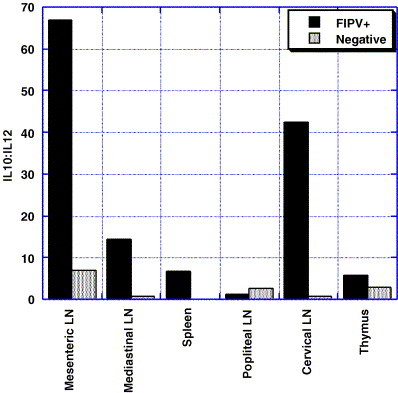
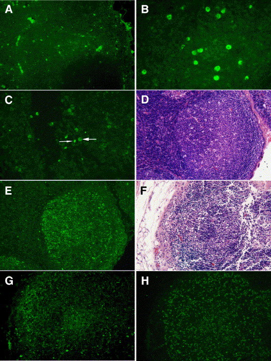
Feline infectious peritonitis virus (FIPV) is a coronavirus that causes sporadic fatal disease in cats characterized by vasculitis, granulomatous inflammation and effusive pleuritis/peritonitis. Histologic changes in lymphoid tissues include lymphoid hyperplasia, lymphoid depletion, histiocytosis, and granuloma formation. Although viremia occurs, histologic lesions are not found uniformly throughout lymphoid tissues. We used experimental infection of cats with a highly pathogenic FIPV isolate, UCD8, to study histologic lesions, virus replication, and cytokine expression in multiple lymphoid tissues during the effusive phase of disease. Viral RNA was found in 76% of central tissues (mediastinal lymph node, spleen, mesenteric lymph node) examined, as compared to 27% of peripheral tissues (popliteal lymph node, cervical lymph node, femoral bone marrow). All tissues positive for virus replication also demonstrated lymphoid depletion. Generally, affected tissues had lower levels of IL-4 and IL-12-p40 mRNA and higher levels of IL-10 mRNA. Although no differences in IFN-gamma or TNF-alpha mRNA were measured, TNF-alpha protein expression was greater in affected tissues and demonstrated a shift in the source of TNF-alpha from macrophages to lymphocytes. Together, these results colocalize FIPV replication, lymphocyte depletion in tissues, and alterations in cytokine transcription and translation. A possible role for TNF-alpha in the previously described FIPV-induced lymphocyte apoptosis is also suggested.
Figures


References
-
- Addie D.D., Toth S., Murray G.D., Jarrett O. The risk of typical and antibody enhanced feline infectious peritonitis among cats from feline coronavirus endemic households. Feline Pract. 1995;23:24–26.
-
- Ayala A., Herdon C.D., Lehman D.L., DeMaso C.M., Ayala C.A., Chaudry I.H. The induction of accelerated thymic programmed cell death during polymicrobial sepsis: control by corticosteroids but not tumor necrosis factor. Shock. 1995;3:259–267. - PubMed
-
- Barlough, J.E., 1988. Feline infectious peritonitis. In: Barlough, J.E. (Ed.), Manual of Small Animal Infectious Diseases. Churchill Livingstone, New York, pp. 63–78.
Publication types
MeSH terms
Substances
LinkOut - more resources
Full Text Sources
Miscellaneous
