

Review
doi: 10.1128/AAC.47.12.3675-3681.2003.
Ribosomal protection proteins and their mechanism of tetracycline resistance
Affiliations
- PMID: 14638464
- PMCID: PMC296194
- DOI: 10.1128/AAC.47.12.3675-3681.2003
Item in Clipboard
Review
Ribosomal protection proteins and their mechanism of tetracycline resistance
Antimicrob Agents Chemother.
2003 Dec.
No abstract available
Figures
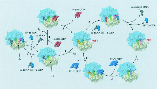
The pathway of Tet(O)-mediated tetracycline release is illustrated by cryo-EM reconstructions of ribosomes in various functional states (2, 45). The natural elongation cycle is represented by reactions a to e, such that if the ribosome is in the posttranslocational state (POST), a ternary complex of EF-Tu-aa-tRNA-GTP can decode the codon presented on the mRNA in the A site (reaction a). After correct codon-anticodon interaction, the GTPase activity of EF-Tu is triggered and the aa-tRNA is accommodated into the A site (reaction b), yielding a pretranslocational ribosome (PRE). After accommodation, the amino group of the A site-bound aa-tRNA attacks the ester bond of the P site-bound peptidyl-tRNA, thereby forming a peptide bond in a reaction called peptidyl transfer (reaction c). Following peptide bond formation, EF-G binds to the ribosome and promotes translocation of the tRNAs from the A and P sites to the P and E sites (reactions d and e), thus completing a single cycle and returning the ribosome to a POST state. Upon tetracycline binding (reaction f), the ribosome allegedly enters a nonproductive cycle illustrated by reactions i and j (4). In this cycle, the ternary complex repeatedly tries to bind aa-tRNA to the A site but fails. Tet(O) is able to rescue the ribosome from this nonproductive cycle by releasing tetracycline from its binding site on the 30S subunit (reaction g). After promoting the release of tetracycline, Tet(O) hydrolyzes its bound GTP and disassociates from the ribosome (reaction h), thereby returning the ribosome to the elongation cycle (reactions a to e). This figure has been reproduced from references and with permission of the publishers.
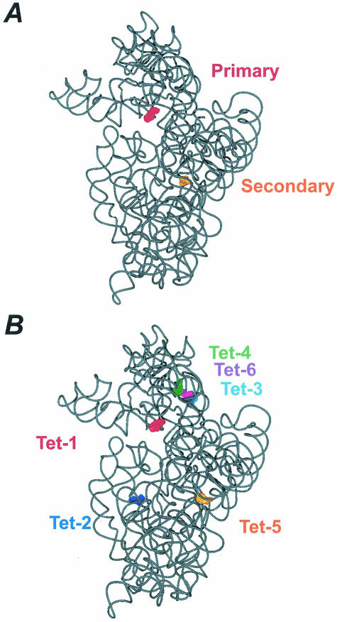
(A) The locations of the tetracycline binding sites determined by Brodersen et al. (4) are shown, where tetracycline bound in the primary site is red (surface representation) and tetracycline bound in the secondary site is orange. The structure shown is derived from the 3.4-Å model (PBD accession no. 1HNW). (B) The locations of the tetracycline binding sites determined by Pioletti et al. (37) are shown, where tetracycline bound to the Tet-1 site is red, tetracycline bound to the Tet-2 site is dark blue, tetracycline bound to the Tet-3 site is cyan, tetracycline bound to the Tet-4 site is green, tetracycline bound to the Tet-5 site is orange, and tetracycline bound to the Tet-6 site is purple. Note, the numbering of the tetracycline binding sites reflects their relative occupancy in the electron density map. The structure shown is derived from the 4.5-Å model (PBD accession no. 1I97). The tetracycline-ribosome interactions in the Tet-1 site are nearly identical to that in the primary site, whereas the Tet-5 and secondary site display distinct interactions. The figures were prepared with VMD (26) and PovRay (www.povray.org ).
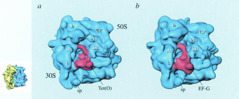
Cryo-EM reconstructions of Tet(O)-GTPγS (45) (a) and EF-G-GMPPCP (1) (b) ribosomal complexes. The ribosome (blue density) is shown in the same orientation as seen in the insert on the left, where the 30S subunit is colored yellow and the 50S subunit is colored blue. Tet(O) and EF-G are shown as red densities. Ribosomal landmarks are indicated. h, head; CP, central protuberance; h38, helix 38 of 23S rRNA; SB, stalk base; sp, spur; sh, shoulder; b, body. This figure has been reproduced from reference with permission of the publisher.
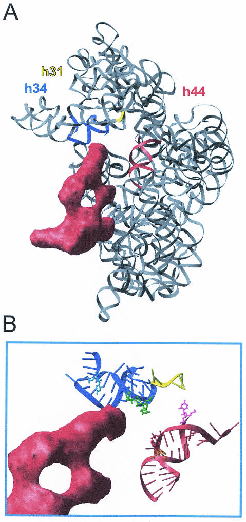
rRNA bases that are altered in DMS modification by the binding of Tet(O) cluster around the decoding center. (A) Tet(O) (red density) (45) bound to the 30S subunit (58) (PDB identification code 1FJF) in the same orientations as seen in panel B. Helices 31 (nucleotides 964 to 968), 34 (nucleotides 1199 to 1217 and 1058 to 1046) and 44 (nucleotides 1400 to 1414 and 1486 to 1503) are represented as yellow, blue, and red ribbons, respectively, and the remaining rRNA is represented as a grey ribbon. (B) Interaction of domain IV of Tet(O) (red density) with the region around the primary tetracycline binding site. Helices 31, 34, and 44 are represented as in panel A. The bases that experience changes in DMS accessibility upon tetracycline (U1052 and C1054, green), EF-G (A1408, orange; C1400, pink), or Tet(O) (C1214, blue; A1408, orange) binding are drawn in a ball and stick representation. This figure has been reproduced from reference with permission of the publisher.
References
-
- Agrawal, R. K., A. B. Heagle, P. Penczek, R. A. Grassucci, and J. Frank. 1999. EF-G-dependent GTP hydrolysis induces translocation accompanied by large conformational changes in the 70S ribosome. Nat. Struct. Biol. 6:643-647. - PubMed
-
- Ban, N., P. Nissen, J. Hansen, P. B. Moore, and T. A. Steitz. 2000. The complete atomic structure of the large ribosomal subunit at 2.4 Å resolution. Science 289:905-920. - PubMed
-
- Brodersen, D. E., W. M. Clemons, A. P. Carter, R. J. Morgan-Warren, B. T. Wimberly, and V. Ramakrishnan. 2000. The structural basis for the action of the antibiotics tetracycline, pactamycin, and hygromycin B on the 30S ribosomal subunit. Cell 103:1143-1154. - PubMed
-
- Burdett, V. 1991. Purification and characterization of Tet(M), a protein that renders ribosomes resistant to tetracycline. J. Biol. Chem. 266:2872-2877. - PubMed
Publication types
MeSH terms
Substances
LinkOut - more resources
Full Text Sources
Other Literature Sources
