. 2003 Dec;71(12):6857-63.
doi: 10.1128/IAI.71.12.6857-6863.2003.

Subtractive hybridization identifies a novel predicted protein mediating epithelial cell invasion by virulent serotype III group B Streptococcus agalactiae

Affiliations

Subtractive hybridization identifies a novel predicted protein mediating epithelial cell invasion by virulent serotype III group B Streptococcus agalactiae

Elisabeth E Adderson et al. Infect Immun. 2003 Dec.

Abstract

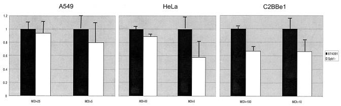
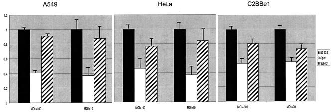
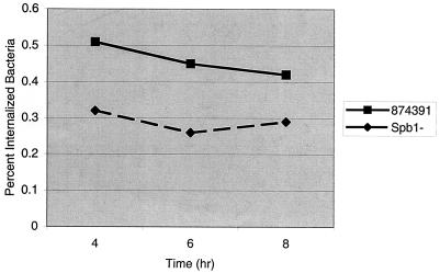
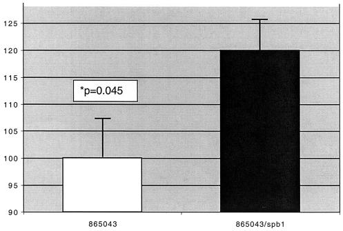
Group B Streptococcus agalactiae bacteria (group B streptococci [GBS]) are the most common cause of serious bacterial infection in newborn infants. The majority of serotype III-related cases of neonatal disease are caused by a genetically related subgroup of bacteria, restriction fragment digest pattern (RDP) type III-3, suggesting that these strains possess unique genes contributing to virulence. We used genomic subtractive hybridization to identify regions of genomic DNA unique to virulent RDP type III-3 GBS strains. Within one of these III-3-specific regions is a 1,506-bp open reading frame, spb1 (surface protein of group B streptococcus 1). A mutant type III GBS strain lacking Spb1 was constructed in virulent RDP type III-3 strain 874391, and the interactions of the wild-type and spb1 isogenic mutant with a variety of epithelial cells important to GBS colonization and infection were compared. While adherence of the spb1 isogenic mutant to A549 respiratory, C2Bbe1 colonic, and HeLa cervical epithelial cells was slightly lower than that of the 874391 strain, invasion of the Spb1(-) mutant was significantly reduced with these cell lines compared to what was seen with 874391. The defect in epithelial invasion was corrected by supplying spb1 in trans. These observations suggest that Spb1 contributes to the pathogenesis of neonatal GBS infection by mediating internalization of virulent serotype III GBS and confirm that understanding of the population structure of bacteria may lead to insights into the pathogenesis of human infections.


Figures

FIG. 1.
FIG. 1.
Nucleic acid and translated amino acid sequences of spb1. Shown are the nucleic acid sequence of the spb1 coding and 5′ noncoding regions and the translated amino acid sequence. A potential ribosomal binding site is underlined with dashes, putative transmembrane domains are underlined with solid lines, an arrow indicates the predicted signal peptide cleavage site, and the LPXTG cell wall-binding domain is shaded. The spb1 nucleotide sequence is available from the GenBank database under accession number AF485279.
FIG. 2.
FIG. 2.
Adherence of Spb1 GBS to epithelial cells. Graphs illustrate the relative adherence of strain Spb1 to A549 respiratory epithelial cells, HeLa cervical epithelial cells, and C2Bbe1 colonic epithelial cells compared to that of RDP type III-3 wild-type strain 874391. Bars represent the mean numbers of adherent bacteria plus standard errors of the mean (SEM) for each inoculum tested and also represent the reduction in invasion of recombinant strains relative to the wild-type strain 874391 for each inoculum tested. Spb1 had lower levels of adhesion than the wild-type strain, but only adherence to C2BBel cells was statistically significant at a MOI of 100 (P < 0.001). Data represent means ± SEM from three to seven separate experiments.
FIG. 3.
FIG. 3.
Invasion of epithelial cells is reduced in Spb1 GBS. Graphs illustrate the relative invasion of A549 respiratory epithelial cells, HeLa cervical epithelial cells, and C2Bbe1 colonic epithelial cells by RDP type III-3 wild-type strain 874391 (black bars), the spb1-deficient isogenic mutant strain Spb1 (white bars), and the Spb1 strain complemented in trans, strain Spb1c (stripped bars). Bars represent the reduction in invasion of recombinant strains relative to that of the wild-type strain 874391 for each inoculum tested. Spb1 invades all epithelial cells significantly less well than the wild-type strain 874391. Complementation of the Spb1 mutant in trans restores invasion to a level comparable to that of the parental strain. Data represent means ± SEM from three or four separate experiments.
FIG. 4.
FIG. 4.
Intracellular persistence of GBS in A549 respiratory epithelial cells. A549 cells were grown to confluence in 24-well tissue culture plates and infected with strain 874391 or Spb1 GBS at a MOI of 5 as described previously. Adherent extracellular bacteria were killed by the addition of tissue culture medium with 5 μg of penicillin/ml and 100 μg of gentamicin/ml followed by incubation at 37°C in 5% CO2. Epithelial cells were lysed at 4, 6, and 8 h after infection, and the percentage of the initial inoculum surviving within cells was quantified. Numbers of intracellular bacteria did not diminish significantly over 8 h. Data represent means from five or six separate experiments.
FIG. 5.
FIG. 5.
Invasion of A549 epithelial cells by GBS strains 865043 and 865043/spb1. The RDP type III-2 strain 865043 was transformed with pMS3545/spb1 to create strain 865043/spb1. Graph illustrates the relative invasion of A549 respiratory epithelial cells by each strain at a MOI of 100. Bars represent the invasion of 865043/spb1 (120.0% ± 7.4%) relative to that of the wild-type strain 865043 (100.0% ± 5.6%). Data represent means ± SEM from five separate experiments.

References

    1. Ausubel, F. M., R. Brent, R. E. Kingston, D. D. Moore, J. G. Seidman, J. A. Smith, K. Struhl, L. M. Albright, D. M. Coen, and A. Varki. 1994. Current protocols in molecular biology. John Wiley & Sons, Inc., New York, N.Y.
    1. Baker, C. J. 1976. Correlation of maternal antibody deficiency with susceptibility to neonatal group B streptococcal infection. N. Engl. J. Med. 294:753-756. - PubMed
    1. Baker, C. J. 2000. Group B streptococcal infections, p. 222-237. In D. L. Stevens, and E. L. Kaplan (ed.), Streptococcal infections: clinical aspects, microbiology, and molecular pathogenesis. Oxford University Press, New York, N.Y.
    1. Baker, C. J., and F. F. Barrett. 1973. Transmission of group B streptococci among parturient women and their neonates. J. Pediatr. 83:919-925. - PubMed
    1. Baker, C. J., and M. S. Edwards. 1995. Group B streptococcal infections, p. 980-1054. In J. Remington, and J. O. Klein (ed.), Infectious diseases of the fetus and newborn infant, 4th ed. W. B. Saunders, Philadelphia, Pa.

Publication types

MeSH terms

Associated data