. 2003 Dec;71(12):7109-18.
doi: 10.1128/IAI.71.12.7109-7118.2003.

Phenotypic switching and mating type switching of Candida glabrata at sites of colonization

Affiliations

Phenotypic switching and mating type switching of Candida glabrata at sites of colonization

Paula J Brockert et al. Infect Immun. 2003 Dec.

Abstract

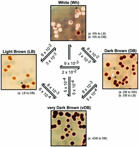
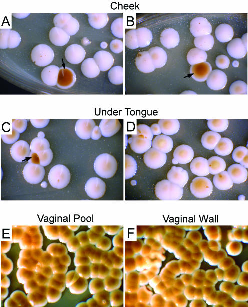
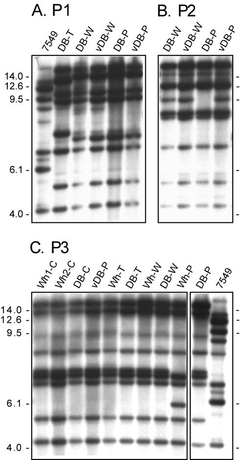
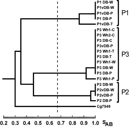
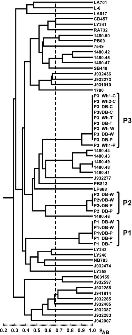
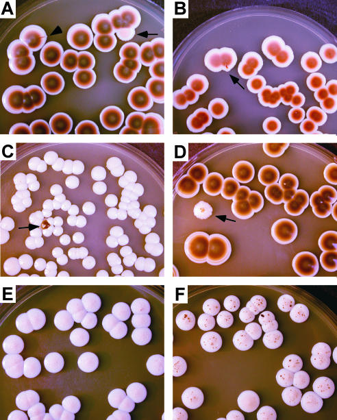
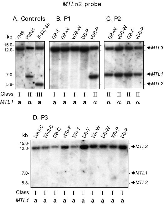
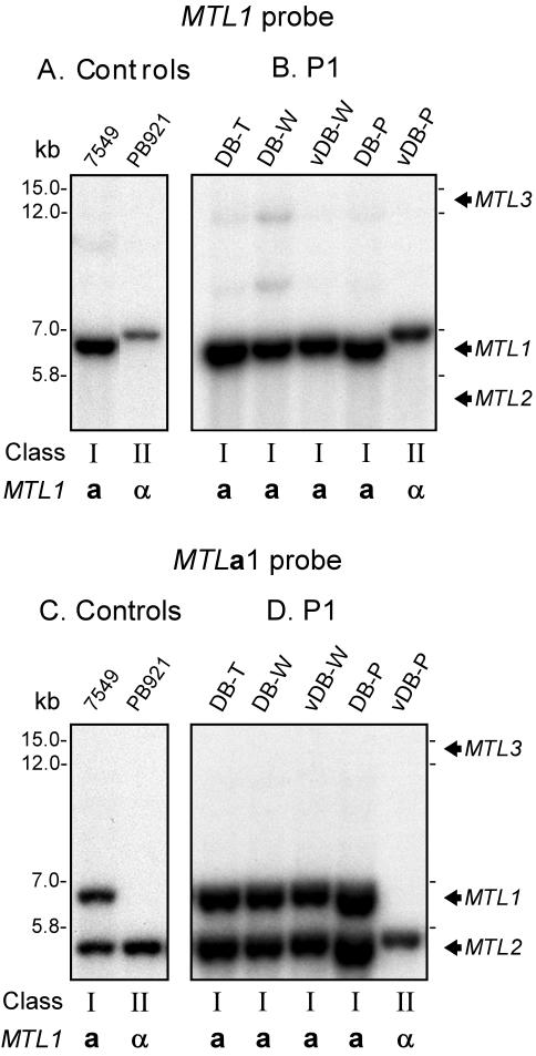
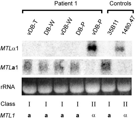
Candida glabrata switches spontaneously at high frequency among the following four graded phenotypes discriminated on agar containing 1 mM CuSO(4): white, light brown, dark brown (DB), and very dark brown. C. glabrata also contains three mating type loci with a configuration similar to that of the Saccharomyces cerevisiae mating type cassette system, suggesting it may also undergo cassette switching at the expression locus MTL1. To analyze both reversible, high-frequency phenotypic switching and mating type switching at sites of colonization, primary samples from the oral cavities and vaginal canals of three patients suffering from C. glabrata vaginitis were clonally plated on agar containing CuSO(4). It was demonstrated that (i) in each vaginitis patient, there was only one colonizing strain; (ii) an individual could have vaginal colonization without oral colonization; (iii) phenotypic switching occurred at sites of colonization; (iv) the DB phenotype predominated at the site of infection in all three patients; (v) genetically unrelated strains switched in similar, but not identical, fashions and caused vaginal infection; (vi) different switch phenotypes of the same strain could simultaneously dominate different body locations in the same host; (vii) pathogenesis could be caused by cells in different mating type classes; and (viii) mating type switching demonstrated at both the genetic and transcription levels occurred in one host.


Figures

FIG. 1.
FIG. 1.
Core switching system in C. glabrata. Cells switch spontaneously among Wh, LB, DB, and vDB phenotypes discriminated on agar containing 1 mM CuSO4. In each box, low-magnification pictures of the dominant phenotypes are presented with examples of switches (a and b in parentheses). The frequencies of variant colony phenotypes are noted on the arrows pointing to those variant phenotypes.
FIG. 2.
FIG. 2.
For patient P3, the Wh phenotype dominated in the oral cavity (cheek and under tongue), while the DB phenotype dominated in the vaginal canal (vaginal wall and vaginal pool). The low-magnification images are of primary colonies plated directly from the original samples (swabs). (A and B) Primary colonies of cheek sample (the arrows point to minority DB colonies). (C and D) Primary colonies of under-tongue sample (the arrow points to a DB colony). (E) Primary colonies of vaginal-pool sample. (F) Primary colonies of vaginal wall sample.
FIG. 3.
FIG. 3.
Southern blot hybridization with the DNA-fingerprinting probe Cg6 demonstrated that only one strain of C. glabrata colonized each patient, that the same strain colonized both the oral cavity and the vaginal canal of each patient, and that the switch phenotypes represented the same strain in each individual. 7549 is a reference strain for normalization in computer-assisted analyses. T, under-tongue sample; C, cheek sample; W, vaginal-wall sample; P, vaginal-pool sample. Wh1 and Wh2 are two independent primary Wh colonies from the same patient. The molecular masses on the left of the blots are in kilodaltons. Note the examples of microevolution in DB-T of patient P1, DB-P of patient P2, and Wh-P of patient P3.
FIG. 4.
FIG. 4.
Dendrogram generated for the DNA-fingerprinted isolates of patients P1, P2, and P3. To generate the dendrogram, the similarity coefficients between all pairs of the DNA-fingerprinted patterns of isolates from patients P1, P2, and P3 were computed. Note that the isolates from each patient form a single cluster, indicating that each patient's collection represented an independent single strain of C. glabrata. See the legend to Fig. 3 for an explanation of the isolate names. The dashed line represents the average SAB computed among all isolate pairs.
FIG. 5.
FIG. 5.
Dendrogram generated for the DNA-fingerprinted isolates from patients P1, P2, and P3 and a large collection of unrelated C. glabrata isolates. Note that even in this larger dendrogram, the clusters of P1, P2, and P3 isolates remain intact, supporting the conclusion that each patient is colonized by a single strain of C. glabrata.The dashed line represents the average SAB computed among all isolate pairs for patients 1, 2, and 3.
FIG. 6.
FIG. 6.
Examples of in vitro switching. Cells from primary colonies were replated (secondary colonies). (A) Colonies from primary DB-VP colonies of patient P1 (the arrowhead points to switch to vDB sector and the arrow points to switch to LB colony). (B) Colonies from primary DB-VP colonies of patient P2 (the arrow points to switch to vDB sector). (C) Colonies from primary Wh-C colonies of patient P3 (the arrow points to switch to DB colony). (D) Colonies from primary DB-VP colonies of patient P3 (the arrow points to switch to Wh). (E) Colonies from primary Wh-C colonies of patient P3 secondary plating incubated for 6 days (note no visible sectoring). (F) Colonies from primary Wh-C colonies of patient P3 secondary plating incubated for 12 days (note sectoring in every colony).
FIG. 7.
FIG. 7.
Southern analysis of mating type classes reveals that infecting strains can be a or α and that mating type switching occurred in patient P1. Southern blots of test isolates from each patient were probed with FuncP2, an antisense oligonucleotide that binds to a unique 5′ end sequence of the MTLα2 ORF. This MTLα2-specific probe discriminates among the three major classes of C. glabrata, class I (a), class II (α), and class III (a) (31). (A) reference strains for the three classes representing the three discriminating patterns. (B) isolates from patient P1. (C) isolates from patient P2. (D) isolates from patient P3. The mating type classes and MTL1 genotypes are provided below the blots. Note that the prevailing patterns of the isolates from patient P1, P2, and P3 are I (a), II (α), and I (a), respectively. Note also the single mating type switch from class I to class II in isolate vDB-P of patient P1. In Fig. 3A, it was demonstrated by Cg6 DNA fingerprinting that the five P1 isolates, including vDB-P, represent the same strain. See the legend to Fig. 3 for an explanation of the isolate names. The positions of MTL1, MTL2, and MTL3 bands are noted to the right of the blots (31).
FIG. 8.
FIG. 8.
Southern blot hybridization analyses with probes for the MTL1 locus and the MTLa1 ORF support the conclusion that a mating type switch is responsible for the vDB-P pattern. (A) Reference strain patterns for class I (a) and class II (α) generated by Southern blot hybridization with a probe that specifically binds to the MTL1a2 3′ flanking sequence (15). (B) Southern blot hybridization patterns of primary P1 isolates with the MTL1a2 3′ flanking sequence probe. (C) Reference strain patterns for classes I (a) and II (α) generated by Southern blot hybridization with the MTLa1 ORF probe. (D) Southern blot hybridization patterns of primary P1 isolates with the MTLa1 ORF probe. Note that in both cases the isolates DB-T, DB-W, vDB-W, and DB-P exhibit the class I (a) pattern while vDB-P exhibits the class II (α) pattern. See the legend to Fig. 3 for explanations of isolate names.
FIG. 9.
FIG. 9.
Northern blot analyses with probes for MTLa1 and MTLα1 transcripts reveal that the genetic switch from a to α at the MTL1 locus in vDB-P was accompanied by a switch from MTLa1 to MTLα1 expression. The expression patterns of the α strain 35B11 and the a strain 1480.47 are presented at the end of the gel for reference. While isolates vDB-T, DB-W, vDB-W, and DP-P, all a at the MTL1 locus, express MTLa1 and not MTLα1, isolate vDB-P, which is α at the MTL1 locus, expresses MTLα1 and not MTLa1. See the legend to Fig. 3 for an explanation of the isolate names.

References

    1. Csank, C., and K. Haynes. 2000. Candida glabrata displays pseudohyphal growth. FEMS Microbiol. Lett. 189:115-120. - PubMed
    1. Fidel, P. L., J. A. Vazquez, and J. D. Sobel. 1999. Candida glabrata: review of epidemiology, pathogenesis and clinical disease with comparison to C. albicans. Clin. Microbiol. Rev. 12:80-96. - PMC - PubMed
    1. Herskowitz, I., J. Rine, and J. N. Strathern. 1992. Mating-type determination and mating-type interconversion in Saccharomyces cerevisiae, p. 583-656. In E. W. Jones, J. R. Pringle, and J. R. Broach (ed.), The molecular and cellular biology of the yeast Saccharomyces. Cold Spring Harbor Laboratory Press, Cold Spring Harbor, N.Y.
    1. Hull, C. M., and A. D. Johnson. 1999. Identification of a mating type-like locus in the asexual pathogenic yeast Candida albicans. Science 285:1271-1275. - PubMed
    1. Kolotila, M. P., and R. D. Diamond. 1990. Effects of neutrophils and in vitro oxidants on survival and phenotypic switching of Candida albicans WO-1. Infect. Immun. 58:1174-1179. - PMC - PubMed

Publication types

LinkOut - more resources