. 2003 Dec;71(12):7119-28.
doi: 10.1128/IAI.71.12.7119-7128.2003.

Recruitment of complement factor H-like protein 1 promotes intracellular invasion by group A streptococci

Affiliations

Recruitment of complement factor H-like protein 1 promotes intracellular invasion by group A streptococci

Vinod Pandiripally et al. Infect Immun. 2003 Dec.

Abstract

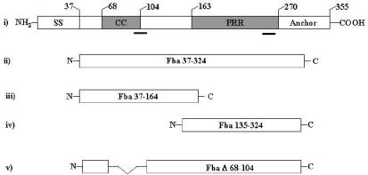
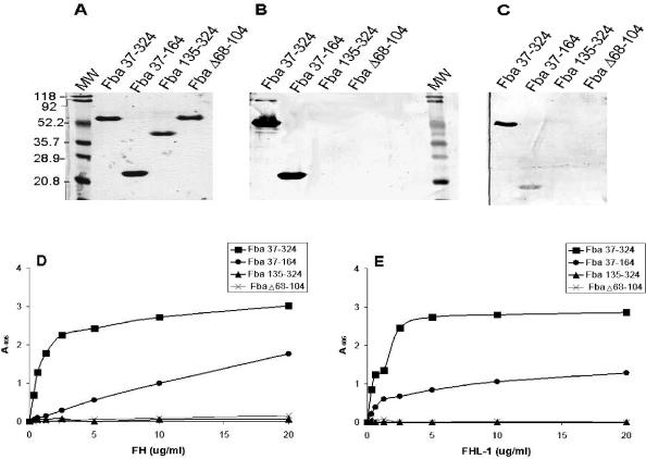
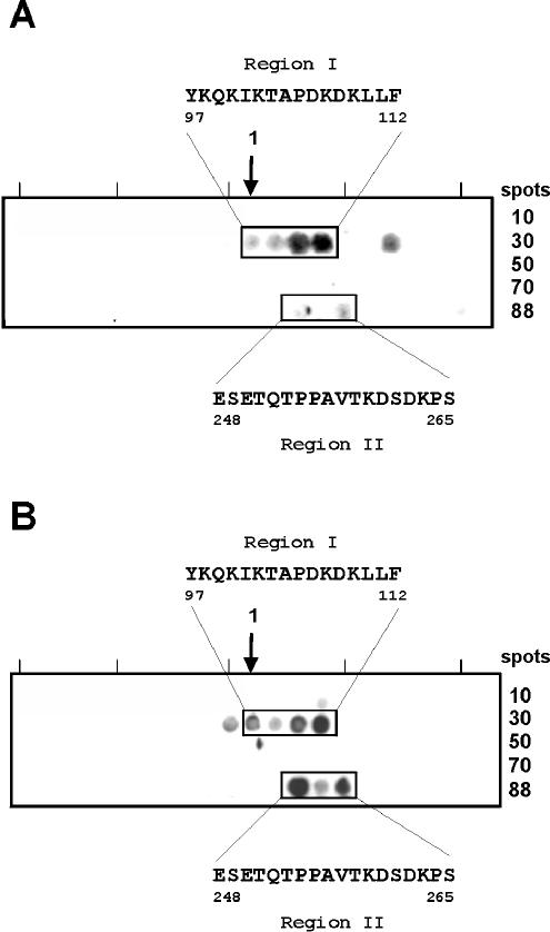
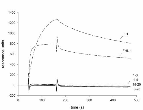
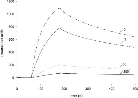
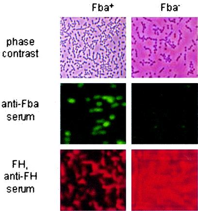
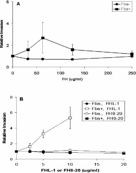
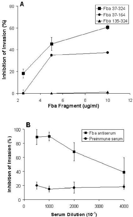
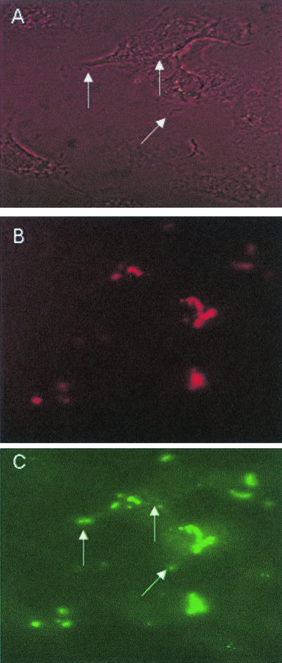
Numerous microbial pathogens exploit complement regulatory proteins such as factor H (FH) and factor H-like protein 1 (FHL-1) for immune evasion. Fba is an FHL-1 and FH binding protein expressed on the surface of the human pathogenic bacterium, Streptococcus pyogenes, a common agent of pharyngeal, skin, and soft-tissue infections. In the present study, we demonstrate that Fba and FHL-1 work in concert to promote invasion of epithelial cells by S. pyogenes. Fba fragments were expressed as recombinant proteins and assayed for binding of FHL-1 and FH by Western blotting, enzyme-linked immunosorbent assay, and surface plasmon resonance. A binding site for FHL-1 and FH was localized to the N-terminal half of Fba, a region predicted to contain a coiled-coil domain. Deletion of this coiled-coil domain greatly reduced FHL-1 and FH binding. PepSpot analyses identified a 16-amino-acid segment of Fba which overlaps the coiled-coil domain that binds both FHL-1 and FH. To localize the Fba binding site in FHL-1 and FH, surface plasmon resonance was used to assess the interactions between the streptococcal protein and a series of recombinant FH deletion constructs. The Fba binding site was localized to short consensus repeat 7 (SCR 7), a domain common to FHL-1 and FH. SCR 7 contains a heparin binding site, and heparin was found to inhibit FHL-1 binding to Fba. FHL-1 promoted entry of Fba(+) group A streptococci into epithelial cells in a dose-dependent manner but did not affect invasion by an isogenic fba mutant. To our knowledge, this is the first report of a bacterial pathogen exploiting a soluble complement regulatory protein for entry into host cells.


Figures

FIG. 1.
FIG. 1.
Recombinant Fba constructs used for this study. Schematic representations of Fba and Fba derivatives are shown. (i) Structural and functional domains of Fba are as follows: SS, signal sequence; CC, coiled-coil region; PRR, proline-rich repeats; anchor, cell wall and membrane anchoring region. Numbers above the figure indicate the terminal amino acid residue of each domain. The bars below the figure indicate amino acid segments shown by PepSpot analyses to bind FHL-1 and FH. (ii to iv) Fba derivatives expressed as recombinant proteins. The numbers within the figures indicate Fba amino acid residues present in each derivative, where residue 1 is the N-terminal formylmethionine (accession number AB040536). (v) FbaΔ68-104 has an internal deletion of amino acid residues 68 to 104.
FIG. 2.
FIG. 2.
Localization of the FHL-1 and FH binding sites in Fba. (A) Coomassie blue-stained SDS-PAGE gel of the recombinant Fba proteins used for this study. The name of each Fba derivative is listed above the gel. MW, molecular mass standards. Numbers to the left of the figure indicate the sizes of each protein standard, in kilodaltons. (B) FH binding by Fba derivatives. The same proteins shown in panel A were transferred to nitrocellulose, blocked, and incubated successively with 10 μg of FH per ml, goat anti-human FH antibody, and an alkaline phosphatase-labeled secondary antibody. No bands were detectable on a duplicate blot incubated with only the primary and secondary antibodies (not shown). At least two independent experiments were performed with each Fba derivative. (C) FHL-1 binding by Fba derivatives. Binding was performed as described for panel B except that the membrane was incubated with FHL-1. (D) FH binding to immobilized Fba. The recombinant Fba derivatives were adsorbed onto microtiter plates and incubated with various concentrations of FH. Binding was detected with FH antiserum and a labeled secondary antibody. Data represent the means from a representative experiment, wherein each assay was performed in triplicate. Each recombinant protein was tested in at least two independent experiments. (E) FHL-1 binding by Fba derivatives. Binding assays were performed as described for panel D except that FHL-1 was substituted for FH. A405, absorbance at 405 nm.
FIG. 3.
FIG. 3.
Mapping of the FHL-1 and FH binding sites in Fba by PepSpot analysis. A total of 88 peptides, spanning the entire Fba protein (excluding the signal sequence and cell anchoring domains), were synthesized and immobilized. Each peptide was 13 amino acid residues in length and had an overlap of 10 amino acids. The membranes were blocked and incubated with FHL-1 (A) or FH (B) and ligand binding was detected with specific antisera. The boxed regions indicate peptides that bound ligand. The amino acid sequences that mediate binding are listed. Numbers refer to amino acid residues of Fba (accession number AB040536), where residue 1 is the N-terminal formylmethionine.
FIG. 4.
FIG. 4.
Localization of the Fba binding site in FHL-1 and FH by the surface plasmon resonance technique. Fba37-324 was coupled to the surfaces of sensor chips, and either FHL-1, FH, or an FH deletion construct, as indicated, was injected into the flow cell. A flow cell that had no protein added was used as a control. Binding was detected as an increase in resonance units over time. Numbers, e.g., 1-6, 1-4, indicate the SCRs represented in each FH deletion construct. A representative sensorgram is shown. Binding experiments were performed three times, with similar results.
FIG. 5.
FIG. 5.
Inhibition of FHL-1 binding to Fba by heparin. Binding assays were performed as described for Fig. 4, except that reactions contained the indicated concentration (3, 30, or 300 μg per ml) of heparin.
FIG. 6.
FIG. 6.
Surface labeling of GAS with Fba antibodies and FH. Fba+ (90-226 emm1::km) and Fba GAS (DC276) were incubated with Fba antiserum (middle) followed by FITC-labeled goat anti-rabbit IgG. Bacteria for the lower panels were incubated with FH, goat anti-human FH serum, and donkey anti-rabbit IgG conjugated to rhodamine red.
FIG. 7.
FIG. 7.
Fba- and FHL-1-dependent invasion of A549 cells. (A) Fba+ (90-226 emm1::km) (squares) and Fba (DC276) (circles) GAS were suspended in RPMI medium or in medium containing various levels of FH and inoculated onto monolayers of A549 cells. The numbers of internalized GAS were determined by a standard antibiotic protection assay, as described in Materials and Methods. For each experiment, the number of CFU recovered from monolayers infected with GAS in the absence of FH was assigned a value of 1.0, and other data are expressed relative to that value. Data are from three independent experiments in which each assay was performed in duplicate. Bars represent standard deviations. (B) Invasion in the presence of various concentrations of FHL-1 (open symbols) or an FH deletion construct that represented SCRs 8 to 20 (filled symbols). Data are presented as described for panel A. The maximum concentrations of FHL-1 and FH8-20 used (10 and 21 μg/ml, respectively) represent equimolar concentrations of the proteins. The overall efficiencies of internalization, in the absence of FHL-1 or FH, were 0.17% ± 0.03% for the Fba+ strain and 0.26% ± 0.12% for the fba mutant strain.
FIG. 8.
FIG. 8.
Inhibition of intracellular invasion by recombinant Fba proteins and Fba antiserum. A549 cells were infected with GAS strain 90-226 emm1::km suspended in RPMI medium containing 15 μg of FHL-1 per ml and various concentrations of either a recombinant Fba derivative (A), Fba antiserum (B), or preimmune serum (B). The numbers of internalized GAS were determined by a standard antibiotic protection assay, as described in Materials and Methods. The percentages of invasion inhibition were calculated relative to those of control wells lacking either Fba or serum. Data are from at least two independent experiments in which each assay was performed in duplicate. Bars represent standard deviations.
FIG. 9.
FIG. 9.
Immunofluorescence microscopy of FHL-1-Fba-mediated invasion. A549 cells were infected with GAS strain 90-226 emm1::km in the presence of FHL-1. After incubation, cells were fixed, blocked, and incubated with rabbit anti-Fba serum, followed by incubation with donkey anti-rabbit IgG labeled with rhodamine red. After removal of unbound antibodies, cells were permeabilized with Triton X-100 and incubated successively with anti-Fba serum and goat anti-rabbit IgG labeled with FITC. Specimens were examined by fluorescence microscopy for intracellular (FITC-labeled) and extracellular (FITC- and rhodamine red-labeled) GAS. (A) Phase-contrast microscopy of infected A549 cells. (B) The same field as that shown in the other panels, irradiated for visualization of extracellular GAS. (C) The same field viewed for the presence of intracellular and extracellular GAS. Arrows in panels A and C indicate intracellular streptococci.

Similar articles

Cited by

References

    1. Areschoug, T., M. Stalhammar-Carlemalm, I. Karlsson, and G. Lindahl. 2002. Streptococcal beta protein has separate binding sites for human factor H and IgA-Fc. J. Biol. Chem. 277:12642-12648. - PubMed
    1. Blackmore, T. K., V. A. Fischetti, T. A. Sadlon, H. M. Ward, and D. L. Gordon. 1998. M protein of the group A streptococcus binds to the seventh short consensus repeat of human complement factor H. Infect. Immun. 66:1427-1431. - PMC - PubMed
    1. Cue, D., P. E. Dombek, and P. P. Cleary. 2000. Intracellular invasion by Streptococcus pyogenes: invasins, host receptors and relevance to human disease, p. 27-33. In V. A. Fischetti et al. (ed.), Gram-positive pathogens. American Society for Microbiology, Washington, D.C. - PMC - PubMed
    1. Cue, D., P. E. Dombek, H. Lam, and P. P. Cleary. 1998. Streptococcus pyogenes serotype M1 encodes multiple pathways for entry into human epithelial cells. Infect. Immun. 66:4593-4601. - PMC - PubMed
    1. Cue, D., S. O. Southern, P. J. Southern, J. Prabhakar, W. Lorelli, J. M. Smallheer, S. A. Mousa, and P. P. Cleary. 2000. A nonpeptide integrin antagonist can inhibit epithelial cell ingestion of Streptococcus pyogenes by blocking formation of integrin α5β1-fibronectin-M1 protein complexes. Proc. Natl. Acad. Sci. USA 97:2858-2863. - PMC - PubMed

Publication types