. 2003 Dec 1;198(11):1753-7.
doi: 10.1084/jem.20031030. Epub 2003 Nov 24.

Vigorous premalignancy-specific effector T cell response in the bone marrow of patients with monoclonal gammopathy

Affiliations

Vigorous premalignancy-specific effector T cell response in the bone marrow of patients with monoclonal gammopathy

Madhav V Dhodapkar et al. J Exp Med. .

Abstract

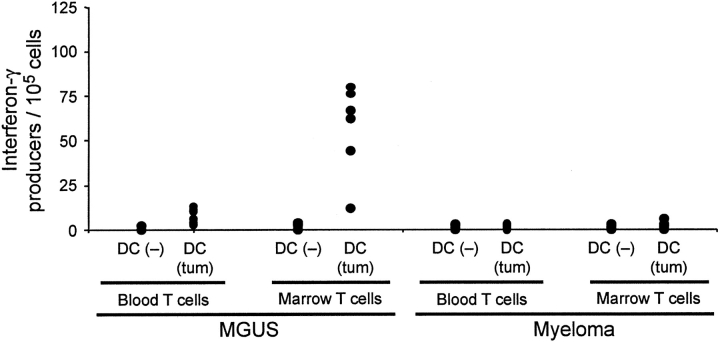
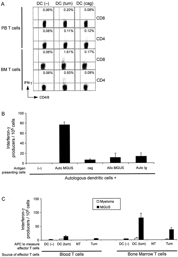
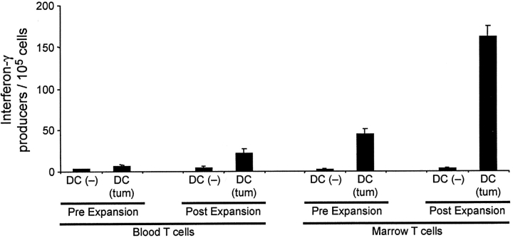
Most approaches targeting the immune system against tumors have focused on patients with established tumors. However, whether the immune system can recognize preneoplastic stages of human cancer is not known. Here we show that patients with preneoplastic gammopathy mount a vigorous T cell response to autologous premalignant cells. This preneoplasia-specific CD4+ and CD8+ T cell response is detected in freshly isolated T cells from the BM. T cells from myeloma marrow lack this tumor-specific rapid effector function. These data provide direct evidence for tumor specific immune recognition in human preneoplasia and suggest a possible role for the immune system in influencing the early growth of transformed cells, long before the development of clinical cancer.


Figures

Figure 1.
Figure 1.
Enrichment of preneoplasia-specific effectors in the BM of MGUS patients. Freshly isolated blood or BM T cells from patients with MGUS (n = 6) or myeloma (n = 6) were tested for the recognition of autologous tumor/preneoplasia-loaded DCs (DC-Tum) or unpulsed DCs (DC) in a 16-h ELISPOT assay for IFN-γ–producing cells.
Figure 2.
Figure 2.
Characterization of preneoplasia-specific immune effectors in the MGUS BM. (A) Preneoplasia-specific IFN-γ–producing T cells consist of both CD4+ and CD8+ T cells. Freshly isolated blood or BM T cells from a patient with MGUS were cultured for 12 h with autologous control DCs, DCs loaded with autologous preneoplastic cells, and DCs loaded with the cag myeloma cell line. IFN-γ–producing CD4+ and CD8+ T cells were examined by intracellular cytokine flow cytometry. Percentages refer to the proportion of CD4+ or CD8+ T cells producing IFN-γ. Data are representative of similar findings on three patients. (B) Preneoplasia-reactive effectors are mostly specific for antigens on autologous preneoplastic cells. BM T cells from a patient with MGUS were tested for the recognition of autologous control DCs, DCs loaded with autologous preneoplastic cells, cag myeloma cell line, preneoplastic cells from a different MGUS patient (allo), or exposed to 10 μg/ml tumor-derived Ig. IFN-γ–producing cells were quantified by a 16-h ELISPOT assay. Data are representative of similar experiments on two patients. (C) Recognition of fresh autologous MGUS tumor cells by immune cells in the BM. Freshly isolated blood or BM T cells from patients with MGUS or myeloma were tested for the recognition of autologous tumor/preneoplasia-loaded DCs (DC-Tum) or unpulsed DCs (DC), or tumor/preneoplasia (Tum; CD138+) or nontumor (NT; CD138−) fractions from the BM in a 16 h ELISPOT assay for IFN-γ–producing cells. Data on the recognition of MGUS tumor cells are representative of findings from two patients.
Figure 3.
Figure 3.
Antipreneoplasia T cells from MGUS patients can be expanded in vitro using autologous preneoplastic cell–loaded DCs. Blood or marrow T cells from a patient with MGUS were cocultured with DCs loaded with autologous MGUS tumor cells. Tumor antigen–specific T cells were quantified by testing reactivity to autologous control DCs or tumor loaded DCs using a 16-h IFN-γ ELISPOT assay. Data shown are representative of similar findings from two patients.

Comment in

Similar articles

Cited by

References

    1. Kyle, R.A., T.M. Therneau, S.V. Rajkumar, J.R. Offord, D.R. Larson, M.F. Plevak, and L.J. Melton, III. 2002. A long-term study of prognosis in monoclonal gammopathy of undetermined significance. N. Engl. J. Med. 346:564–569. - PubMed
    1. Fonseca, R., R.J. Bailey, G.J. Ahmann, S.V. Rajkumar, J.D. Hoyer, J.A. Lust, R.A. Kyle, M.A. Gertz, P.R. Greipp, and G.W. Dewald. 2002. Genomic abnormalities in monoclonal gammopathy of undetermined significance. Blood. 100:1417–1424. - PubMed
    1. Zhan, F., J. Hardin, B. Kordsmeier, K. Bumm, M. Zheng, E. Tian, R. Sanderson, Y. Yang, C. Wilson, M. Zangari, et al. 2002. Global gene expression profiling of multiple myeloma, monoclonal gammopathy of undetermined significance, and normal bone marrow plasma cells. Blood. 99:1745–1757. - PubMed
    1. Kuehl, W.M., and P.L. Bergsagel. 2002. Multiple myeloma: evolving genetic events and host interactions. Nat. Rev. Cancer. 2:175–187. - PubMed
    1. Dhodapkar, M.V., J. Krasovsky, and K. Olson. 2002. T cells from the tumor microenvironment of patients with progressive myeloma can generate strong tumor specific cytolytic responses to autologous tumor loaded dendritic cells. Proc. Natl. Acad. Sci. USA. 99:13009–13013. - PMC - PubMed

Publication types

MeSH terms

Substances