. 2003 Nov 24;163(4):743-53.
doi: 10.1083/jcb.200308020.

Distinct conformations of the kinesin Unc104 neck regulate a monomer to dimer motor transition

Affiliations

Distinct conformations of the kinesin Unc104 neck regulate a monomer to dimer motor transition

Jawdat Al-Bassam et al. J Cell Biol. .

Abstract

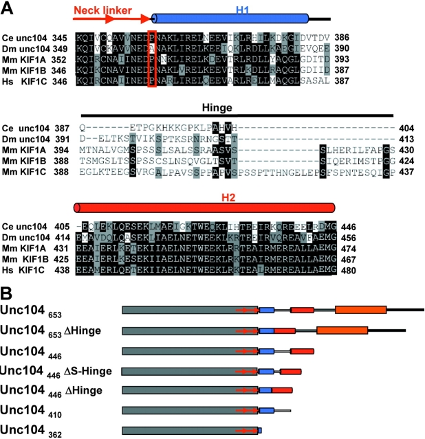
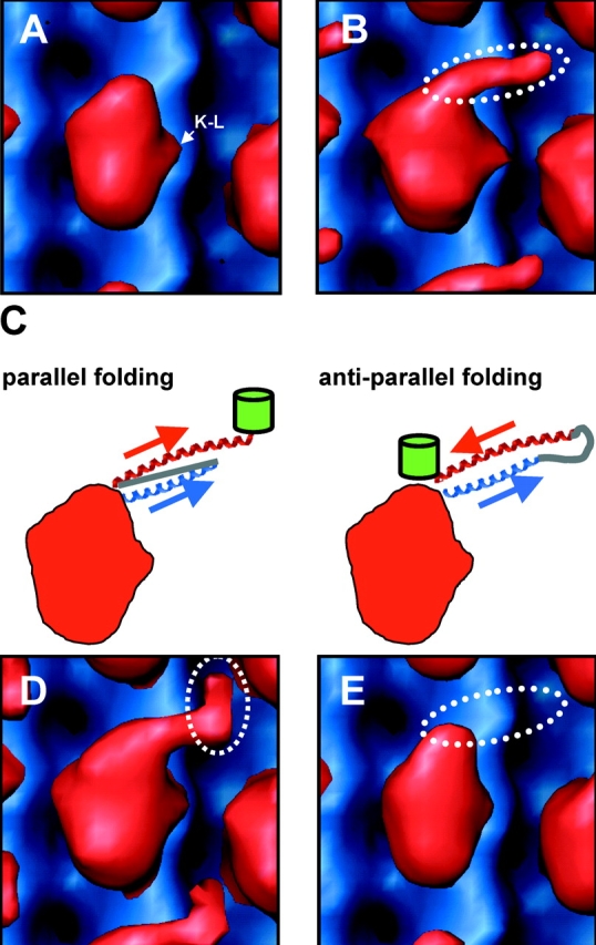
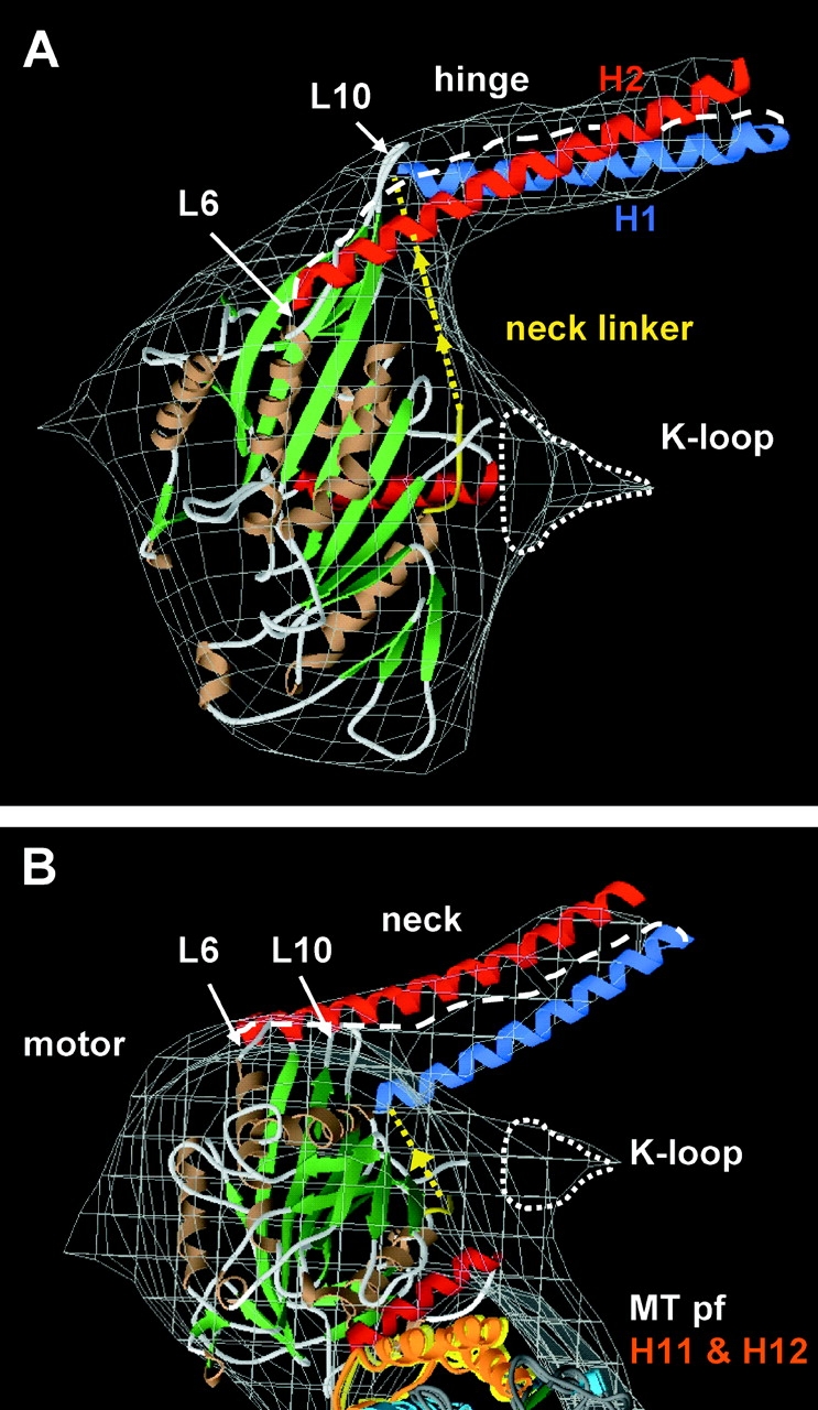
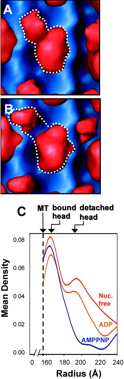
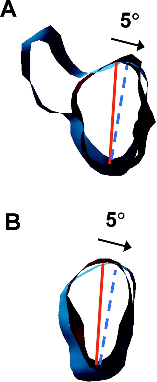
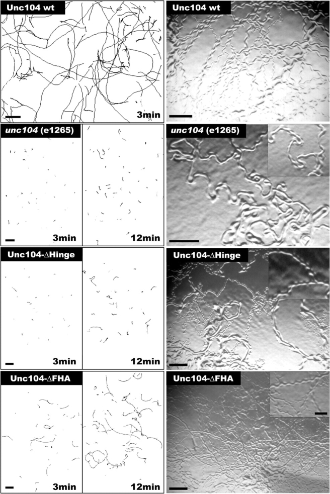
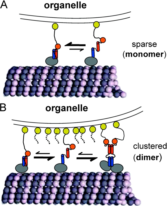
Caenhorhabditis elegans Unc104 kinesin transports synaptic vesicles at rapid velocities. Unc104 is primarily monomeric in solution, but recent motility studies suggest that it may dimerize when concentrated on membranes. Using cryo-electron microscopy, we observe two conformations of microtubule-bound Unc104: a monomeric state in which the two neck helices form an intramolecular, parallel coiled coil; and a dimeric state in which the neck helices form an intermolecular coiled coil. The intramolecular folded conformation is abolished by deletion of a flexible hinge separating the neck helices, indicating that it acts as a spacer to accommodate the parallel coiled-coil configuration. The neck hinge deletion mutation does not alter motor velocity in vitro but produces a severe uncoordinated phenotype in transgenic C. elegans, suggesting that the folded conformation plays an important role in motor regulation. We suggest that the Unc104 neck regulates motility by switching from a self-folded, repressed state to a dimerized conformation that can support fast processive movement.


Figures

Figure 1.
Figure 1.
Sequence comparisons and Unc104 constructs used in the paper. (A) Sequences from C. elegans (Ce) Unc104, D. Melanogaster (Dm) Unc104, M. musculus (Mm) KIF1A and KIF1B, and H. sapiens (Hs) KIF1C were compared using Clustal W (Aiyar, 2000). The Unc104 neck linker is class-conserved and ends with a conserved proline (red box). The neck consists of two predicted α-helical segments, H1 and H2, separated by a nonconserved hinge. Helical prediction was done with PHDsec (Rost and Sander, 1994). H1 consists of ∼26 residues (blue cylinder) after the neck linker. The hinge (solid line) consists of 18–49 residues. H2 consists of ∼41 residues (red cylinder). Conserved residues are shaded black, semi-conserved residues are gray. (B) Schematic representation of the Unc104 constructs used in this paper. Domains are indicated as follows: catalytic core, gray; neck linker, red arrows; H1, blue cylinder; hinge, unshaded line; H2, red cylinder; neck-FHA linker, gray line; FHA domain, orange, sequence after FHA, black line.
Figure 2.
Figure 2.
Structure of the Unc104 neck. Front view (A) of molecular surface representation the Unc104362-AMPPNP map (red) aligned and displayed with an undecorated microtubule map (blue). Density attributable to the K-loop is indicated (K–L). (B) The same view of the Unc104446-AMPPNP map. Additional density projecting from the tip of the catalytic core is attributable to the Unc104 neck (dotted ellipse). Possible self-folding models for the Unc104 neck are shown in C. In a parallel folding model, the H1 helix (blue) extends away from the catalytic core and the hinge (gray line) backtracks to allow H2 to lie parallel to H1. In the antiparallel folding model, the hinge lies distally and H2 backtracks in antiparallel fashion along H1. The green barrels represent the expected location of the GFP in the two models. The Unc104446-GFP map is shown in D. Additional density is located at the distal end of the neck density, consistent with the parallel folding model. The Unc104446-ΔHinge map is shown in E. When the hinge is absent, the neck density is not visualized, again consistent with the parallel folding model.
Figure 3.
Figure 3.
Modeling of the self-folded Unc104446 neck in the AMPPNP state. (A) Front and (B) top views (from the minus end) of a model in which the KIF1A-AMPPCP and tubulin structures were manually docked into the Unc104446 AMPPNP map (gray wire frame). A model for the ∼26 residue H1 (blue) and the ∼41 residue H2 (red) was constructed from the parallel coiled-coil cortexillin structure (Burkhard et al., 2000). A docked neck linker (yellow) joins the α-helical neck with the catalytic core. The α helices and β sheets in the motor and microtubule are shown in brown and green, respectively. There is additional density (enclosed by a dotted line) near L12 of the KIF1A crystal structure that accounts for the position of the K-loop, as seen previously in KIF1A (Kikkawa et al., 2000). The 45-Å long neck density fits the length of the ∼26-residue H1 α helix. The hinge (white dashed line) connects H1 to H2. As H2 is ∼25 Å longer than H1, and a COOH-terminal GFP is found at the end of the neck density, the NH2-terminal part of H2 must lie over the catalytic core, possibly interacting with loops 6 and 10 (L6 and L10) or associated secondary structure elements at the tip of the catalytic core. (B) In the top view, parts of the two tubulin COOH-terminal helices, H11 and H12 (orange) are closely associated with the catalytic core.
Figure 4.
Figure 4.
Unc104446 dimerizes in ADP and nucleotide-free states. In the(A) ADP and (B) nucleotide-free states, additional density is seen near the clearly recognizable bound head. The density is too large to be the neck and somewhat smaller than expected for a second rigidly attached catalytic core. The appearance is consistent with there being a second catalytic core that is loosely associated with the microtubule-bound catalytic core. Note that the finger density seen in Fig. 2 B is absent, and the detached catalytic core seems to occupy a slightly different position in the two states. (A and B) The dotted lines outline density attributable to the microtubule-bound Unc104 dimer. The MRDDs in the 3D-maps (C) clearly show a peak at a radius of ∼200 Å that is attributable to a second, microtubule-detached, catalytic core in the ADP (orange) and nucleotide-free (red) states. No evidence for a second catalytic core is seen in AMPPNP (blue).
Figure 5.
Figure 5.
The Unc104 catalytic core rotates by5° during the ATPase cycle. Thin slices of the catalytic cores of (A) Unc104446 and (B) Unc104362 in the AMPPNP (red) and ADP (blue) maps superimposed to show the small ∼5° rotation between these two nucleotide states. Solid red and dotted blue lines indicate the approximate orientation of the long axes of the catalytic core in the two states. The arrows indicate the rotational relationship between the Unc104 catalytic cores in the two nucleotide states.
Figure 6.
Figure 6.
Effect of hinge and FHA domain deletions on Unc104 performance in living C. elegans . Comparison of velocities and coordinated movement of unc104 null (e1265) worms with worms expressing wild-type Unc104 wt, Unc104-ΔHinge, or Unc104-ΔFHA constructs. (Left) Worm movement was recorded over 3- or 12-min intervals and the position of the worms was tracked and plotted. Average velocities are 12.7 ± 3.6 mm/min for Unc104-wt (n = 454), 1.1 ± 0.9 mm/min for unc104 null (n = 150), 1.6 ± 1.5 mm/min for Unc104-ΔHinge (n = 419), and 2.1 ± 1.1 mm/min Unc104-ΔFHA (n = 380). The differences in velocity between the null, Unc104-ΔHinge, and Unc104-ΔFHA animals are statistically significant (P < 0.0001). Bars, 2.4 mm. (Right) Worm tracks on an agar plate reveal the degree of coordination. Although Unc104 wt animals move steadily in a sinusoidal motion in one direction over several body lengths, unc104 null worms rarely show coordinated movement. Although Unc104-ΔHinge worms move slowly, their tracks show a slight improvement of coordination compared to unc104 null (inset). Unc104-ΔFHA worms show sinusoidal tracks that are directional over several amplitudes. Bars: 0.5 mm; (inset) 0.2 mm.
Figure 7.
Figure 7.
Model for regulation of monomer to dimer transition by self-folding of the neck helices. When Unc104 is sparsely distributed on an organelle membrane (A), the self-folded state is favored over the unfolded state in a dynamic equilibrium. The self-folded Unc104 monomers may move very slowly and nonprocessively, like Unc104362 (Tomishige et al., 2002). These monomers may show some plus end–directed biased diffusion, but will not generate significant force against an opposing load (Okada et al., 2003). However, when Unc104 is clustered, possibly in lipid rafts, on the organelle membrane at high local concentrations (B), unfolded monomers are recruited into dimers shifting the equilibrium away from the monomeric state. The dimerized Unc104 would then undergo fast processive motility along microtubules and generate maximal force. The monomer to dimer equilibrium shift may occur in solution, but may also be favored by the presence of microtubules. Blue and red cylinders represent H1 and H2, respectively. Orange spheres represent the FHA domains. Yellow spheres represent the membrane PH domain.

Comment in

  • Breathing down the neck of Unc104.
    Wordeman L. Wordeman L. J Cell Biol. 2003 Nov 24;163(4):693-5. doi: 10.1083/jcb.200310137. J Cell Biol. 2003. PMID: 14638852 Free PMC article. Review.

References

    1. Aiyar, A. 2000. The use of CLUSTAL W and CLUSTAL X for multiple sequence alignment. Methods Mol. Biol. 132:221–241. - PubMed
    1. Al-Bassam, J., R.S. Ozer, D. Safer, S. Halpain, and R.A. Milligan. 2002. MAP2 and tau bind longitudinally along the outer ridges of microtubule protofilaments. J. Cell Biol. 157:1187–1196. - PMC - PubMed
    1. Bloom, G.S. 2001. The UNC-104/KIF1 family of kinesins. Curr. Opin. Cell Biol. 13:36–40. - PubMed
    1. Brenner, S. 1974. The genetics of Caenorhabditis elegans. Genetics. 77:71–94. - PMC - PubMed
    1. Burkhard, P., R.A. Kammerer, M.O. Steinmetz, G.P. Bourenkov, and U. Aebi. 2000. The coiled coil trigger site of the rod domain of cortexillin I unveils a distinct network of interhelical and intrahelical salt bridges. Structure Fold. Des. 8:223–230. - PubMed

Publication types

MeSH terms