. 2003 Nov 24;163(4):755-66.
doi: 10.1083/jcb.200308038.

Mammalian GGAs act together to sort mannose 6-phosphate receptors

Affiliations

Mammalian GGAs act together to sort mannose 6-phosphate receptors

Pradipta Ghosh et al. J Cell Biol. .

Abstract

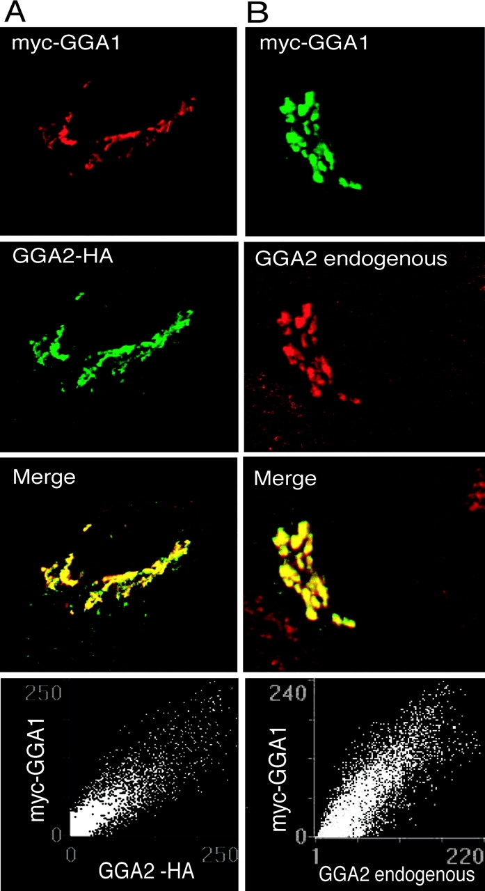
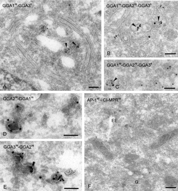
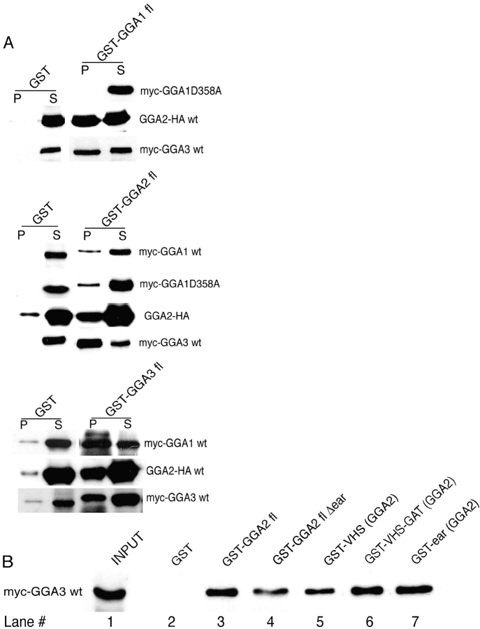
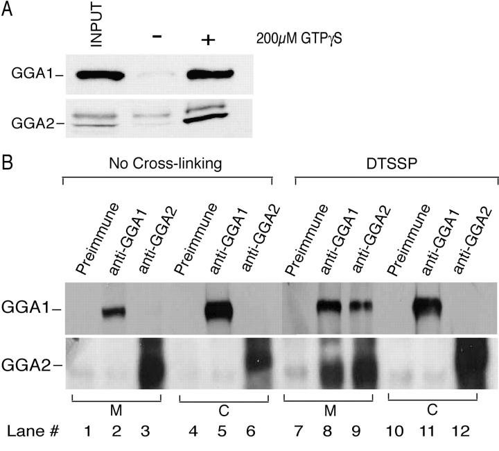
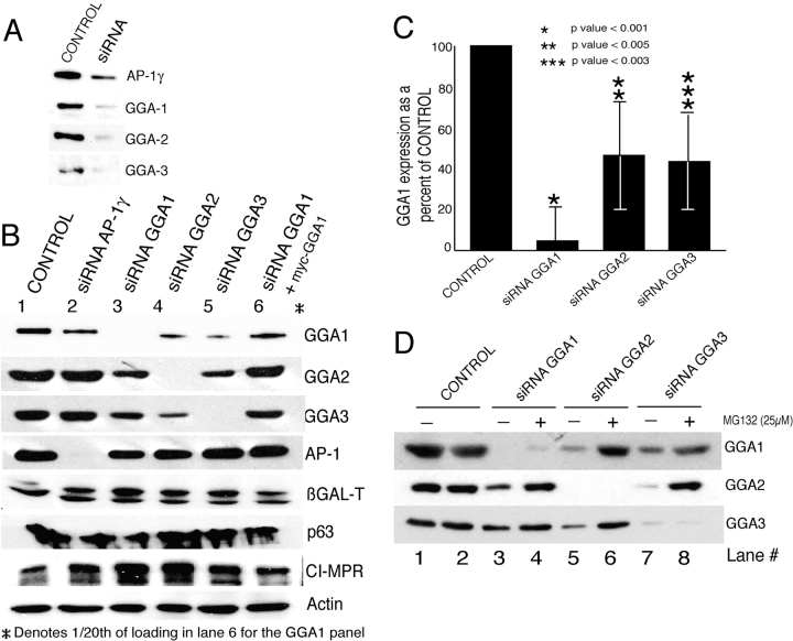
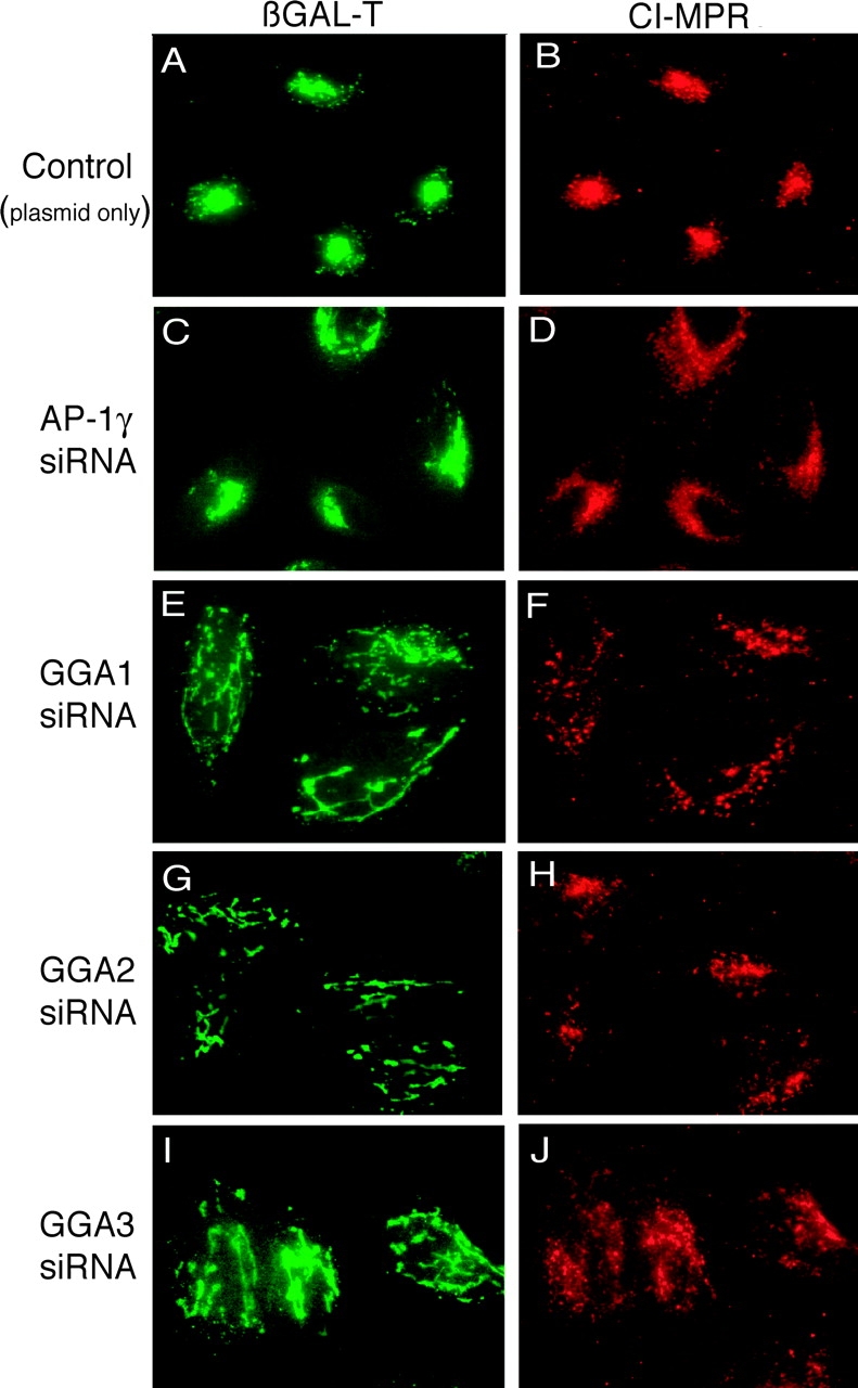
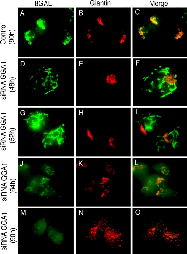
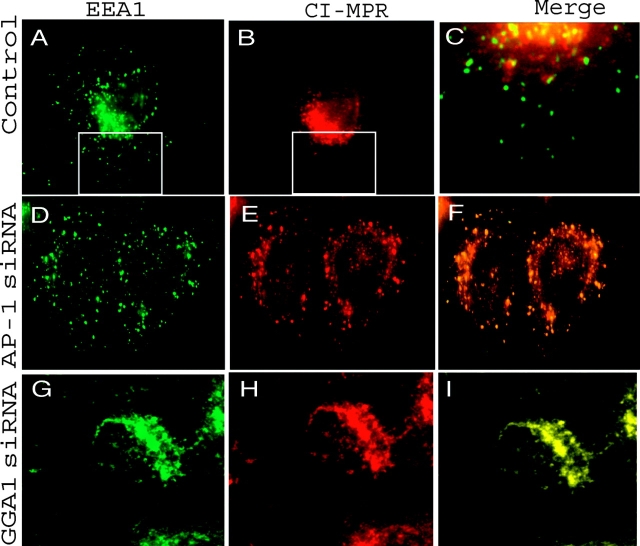
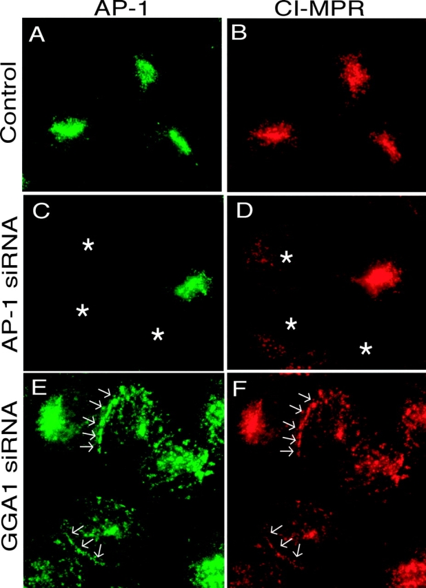
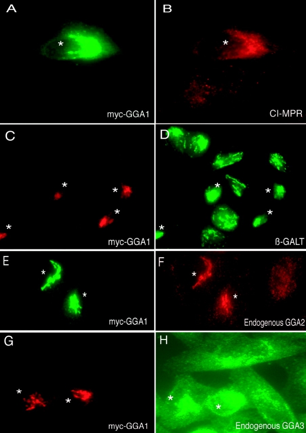
The GGAs (Golgi-localized, gamma ear-containing, ADP ribosylation factor-binding proteins) are multidomain proteins implicated in protein trafficking between the Golgi and endosomes. We examined whether the three mammalian GGAs act independently or together to mediate their functions. Using cryo-immunogold electron microscopy, the three GGAs were shown to colocalize within coated buds and vesicles at the trans-Golgi network (TGN) of HeLa cells. In vitro binding experiments revealed multidomain interactions between the GGAs, and chemical cross-linking experiments demonstrated that GGAs 1 and 2 form a complex on Golgi membranes. RNA interference of each GGA resulted in decreased levels of the other GGAs and their redistribution from the TGN to cytosol. This was associated with impaired incorporation of the cation-independent mannose 6-phosphate receptor into clathrin-coated vesicles at the TGN, partial redistribution of the receptor to endosomes, and missorting of cathepsin D. The morphology of the TGN was also altered. These findings indicate that the three mammalian GGAs cooperate to sort cargo and are required for maintenance of TGN structure.


Figures

Figure 1.
Figure 1.
GGAs 1 and 2 colocalize on the TGN. (A) HeLa cells cotransfected with plasmid DNA encoding myc-GGA1 and GGA2-HA were fixed 48 h after transfection, and double-labeled confocal immunofluorescence was performed using anti-c-myc to detect myc-GGA1 (red) and anti-HA to detect GGA2-HA (green). Confocal images were analyzed and merged using software programs as described in Materials and methods. Scattergram of red and green pixels were plotted on a graph to obtain the colocalization coefficient between myc-GGA1 and GGA2-HA. (B) HeLa cells transfected with plasmid DNA encoding myc-GGA1 were fixed 48 h after transfection and processed for confocal immunofluorescence using anti-c-myc to detect myc-GGA1 (green) and rabbit polyclonal anti-GGA2 antibody to detect endogenous GGA2 (red). A scattergram was plotted as in A to look for colocalization of the red and green pixels.
Figure 2.
Figure 2.
GGAs 1, 2 and 3 colocalize within the same coated buds at the TGN. Electron micrographs of immunogold-labeled cryosections showing the colocalization of GGAs (A–E) and the distribution of CI-MPR and AP-1 in the trans-Golgi area of a GGA1 siRNA cell (F). (A–C) HeLa cells expressing myc-tagged GGA1. Using antibodies against myc, GGA2, and GGA3, double- and triple-labeling experiments with 5-, 10-, and 15-nm gold show that all three GGAs colocalize to coated pits and vesicles at the TGN (arrowheads). D and E show the colocalization of endogenous GGA1, 2, and 3 at coated buds on the TGN in HeLa cells. (F) AP-1 and CI-MPR double immunolabeling in a GGA1-depleted HeLa cell. G, Golgi stack; EE, early endosome. Bars: (A–E) 100 nm, (F) 250 nm.
Figure 3.
Figure 3.
Direct interaction of GGAs with each other in in vitro assays. (A) GST-fused full-length GGA1, GGA2, and GGA3 proteins prebound to glutathione-Sepharose beads were used in binding assays with affinity-purified myc-GGA1wt, myc-GGA1 D358A, myc-GGA3, and GGA2-HA as described in Materials and methods. GST alone was used as a negative control. The bound proteins were eluted by boiling in SDS sample buffer and were subjected to SDS-PAGE and Western blotting using anti-c-myc or anti-HA mAbs to detect GGAs1/3 and GGA2, respectively. P, pellet; S, supernatant. (B) GST-fused GGA2 proteins (full-length, GST-GGA2flΔear, GST-VHS, GST-VHS+GAT, and GST-ear; see Materials and methods for aa sequences) prebound to glutathione-Sepharose beads were used in binding assays with affinity-purified myc-GGA3 wild type. The bound GGA3 was eluted in SDS sample buffer, and an aliquot (20%) of the same was subjected to SDS-PAGE and Western blotting with anti-c-myc mouse mAb. The above result is one of two independent experiments.
Figure 4.
Figure 4.
GGAs form a complex upon recruitment onto Golgi-enriched membranes. (A) Bovine adrenal cytosolic GGAs were recruited onto rat Golgi-enriched membranes in the presence or absence of GTPγS as described in Materials and methods. The membrane-associated proteins were boiled in SDS sample buffer and subjected to SDS-PAGE and Western blotting with either anti-GGA1 or anti-GGA2 pAbs. (B) Recruitment reactions were scaled up and performed as above, followed by chemical cross-linking of membrane-associated proteins with DTSSP as described in Materials and methods. The cross-linked proteins were then isolated by solubilizing the membranes with detergent and were coimmunoprecipitated using either rabbit preimmune sera (negative control) or anti-GGA1 or GGA2 affinity-purified antibodies. Immunoprecipitated proteins were subjected to SDS-PAGE and Western blotting to detect GGA1 and GGA2. A 2-mg aliquot of cytosol (supernatants from the recruitment reactions) was also subjected to cross-linking and coimmunoprecipitation as described in the case of membranes. M, membrane; C, cytosol. Of note, GGA1 migrated slower than expected for its calculated molecular weight.
Figure 5.
Figure 5.
Post-transcriptional silencing of GGAs 1, 2, and 3. (A) HeLa cells transfected with either empty vector (control) or plasmid DNA encoding antisense siRNAs targeting the three GGAs or AP-1γ were harvested 56 h after transfection, and equal aliquots (25 μg) of the cell extracts were subjected to SDS-PAGE and Western blotting with appropriate antibodies to evaluate the efficiency of knockdown achieved. (B) 25-μg Aliquots of cell extracts prepared as in the above experiment were subjected to SDS-PAGE and Western blotting to detect the three GGAs, AP-1, and various marker proteins as indicated in the figure. For lane 6, HeLa cells were cotransfected with GGA1 siRNA plasmid and RNAi-resistant myc-GGA1 as described in Materials and methods. Loading in the first panel of lane 6 was 1/20th of the other lanes because the myc-GGA1 was greatly overexpressed. (C) Level of expression of GGA1 in control cells (plasmid only), GGA1 siRNA, GGA2 siRNA, and GGA3 siRNA cells was detected by Western blotting of cell extracts and was quantitated using a Kodak band densitometer. The amount of GGA1 in each of the cell lines was expressed as a percentage of the control. Error bars represent the values obtained from six independent sets of experiments. P-values were calculated using Sigma Plot. (D) HeLa cells transfected with either vector alone (control) or various siRNA GGA plasmids were subjected to treatment with 25 μM MG132 in DMSO (+) or with DMSO alone (−) for 10 h before harvest. Equal aliquots (33 μg) of cell extracts were subjected to SDS-PAGE and Western blotting to detect the levels of expression of the three GGA proteins.
Figure 6.
Figure 6.
Morphology of GGA1 knockdown cells. HeLa cells were transfected with plasmid DNA encoding either vector only (A and B; control) or siRNAs directed against AP-1γ (C and D), GGA1 (E and F), GGA2 (G and H), and GGA3 (I and J) as described in Materials and methods. Cells were harvested 56 h after transfection and processed for double-labeled immunofluorescence with anti-β-GalT (green) and anti-CI-MPR (red).
Figure 7.
Figure 7.
Disruption of Golgi architecture in GGA knockdown cells. HeLa cells transfected with plasmid DNA expressing GGA1 siRNA were harvested at the indicated times after transfection and processed for double-labeled immunofluorescence with antibodies against β-GalT (green) and giantin (red).
Figure 8.
Figure 8.
Redistribution of CI-MPR to peripheral EEA1-positive compartments. HeLa cells transfected with AP-1 siRNA or GGA1 siRNA were double labeled with anti-CI-MPR (red) and anti-EEA1 (green). C is a blow-up of the boxed areas in A and B.
Figure 9.
Figure 9.
AP-1 and CI-MPR colocalize within peripheral structures. HeLa cells transfected with vector alone (A and B; control), AP-1 siRNA (C and D) and GGA1 siRNA (E and F) were harvested 56 h after transfection and immunostained for AP-1 (green) and CI-MPR (red). Asterisks designate cells with AP-1 knockdown. Arrows highlight areas where AP-1 and the CI-MPR colocalize on tubular processes.
Figure 10.
Figure 10.
Prevention of morphological changes in GGA1 knockdown cells by transfection of RNAi-resistant myc-GGA1. HeLa cells were cotransfected with GGA1 siRNA and RNAi-resistant myc-GGA1 as described in Materials and methods. 56 h after transfection, the cells were harvested for double-labeled immunofluorescence to detect myc-GGA1 (green) and CI-MPR (red; A and B); myc-GGA1 (red) and β-GalT (green; C and D); myc-GGA1 (green) and endogenous GGA2 (red; E and F); and myc-GGA1 (red) and endogenous GGA3 (green; G and H). Asterisks indicate the cells expressing transfected myc-GGA1.

Similar articles

Cited by

References

    1. Black, M.W., and H.R. Pelham. 2000. A selective transport route from Golgi to late endosomes that requires the yeast GGA proteins. J. Cell Biol. 151:587–600. - PMC - PubMed
    1. Boman, A.L., C. Zhang, X. Zhu, and R.A. Kahn. 2000. A family of ADP-ribosylation factor effectors that can alter membrane transport through the trans-Golgi. Mol. Biol. Cell. 11:1241–1255. - PMC - PubMed
    1. Brummelkamp, T.R., R. Bernards, and R. Agami. 2002. A system for stable expression of short interfering RNAs in mammalian cells. Science. 296:550–553. - PubMed
    1. Crump, C.M., Y. Xiang, L. Thomas, F. Gu, C. Austin, S.A. Tooze, and G. Thomas. 2001. PACS-1 binding to adaptors is required for acidic cluster motif-mediated protein traffic. EMBO J. 20:2191–2201. - PMC - PubMed
    1. Dell'Angelica, E.C., R. Puertollano, C. Mullins, R.C. Aguilar, J.D. Vargas, L.M. Hartnell, and J.S. Bonifacino. 2000. GGAs: a family of ADP ribosylation factor-binding proteins related to adaptors and associated with the Golgi complex. J. Cell Biol. 149:81–94. - PMC - PubMed

Publication types

MeSH terms