. 2003 Nov 24;163(4):859-69.
doi: 10.1083/jcb.200304153.

The stromal cell-derived factor-1alpha/CXCR4 ligand-receptor axis is critical for progenitor survival and migration in the pancreas

Affiliations

The stromal cell-derived factor-1alpha/CXCR4 ligand-receptor axis is critical for progenitor survival and migration in the pancreas

Ayse G Kayali et al. J Cell Biol. .

Abstract

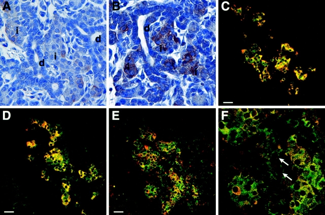
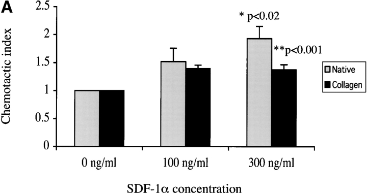
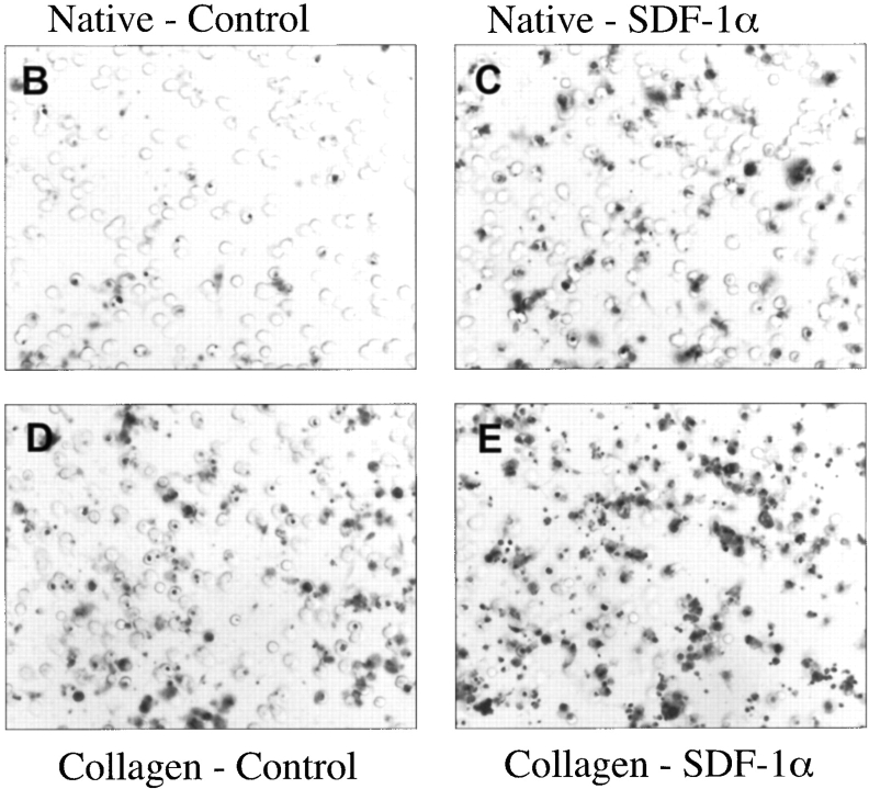
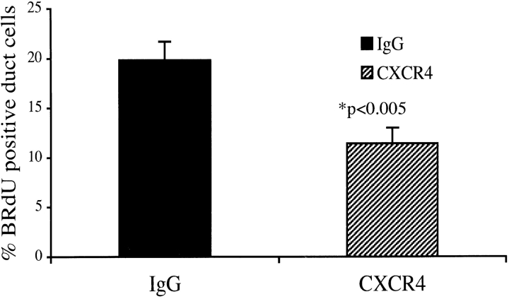
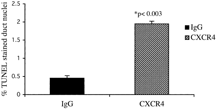
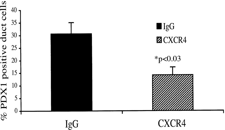
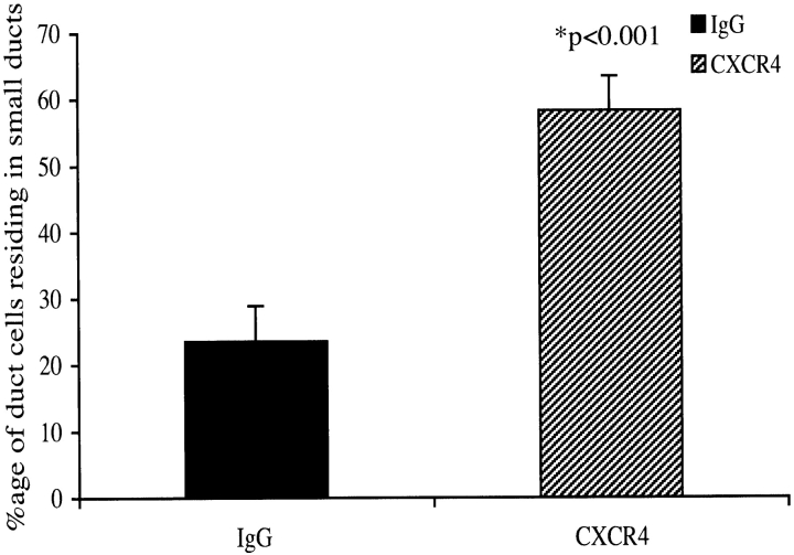
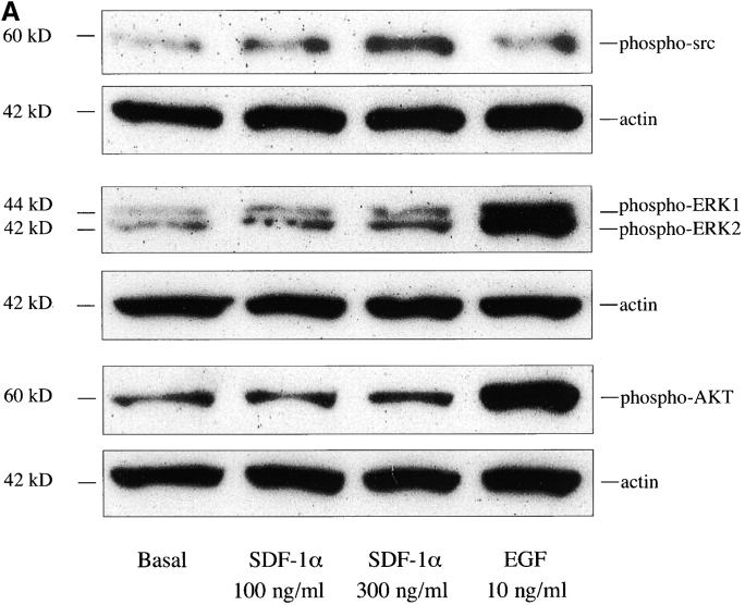
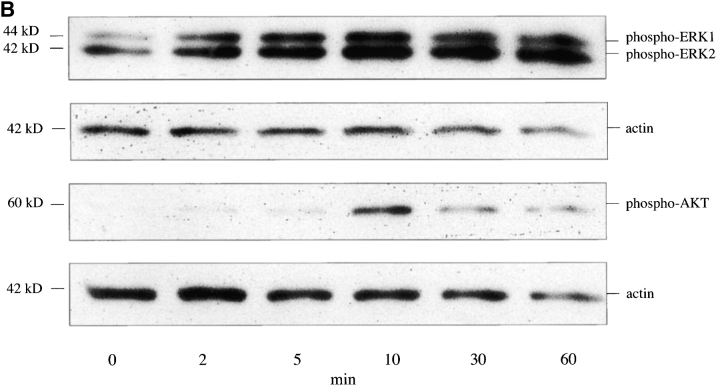
The SDF-1alpha/CXCR4 ligand/chemokine receptor pair is required for appropriate patterning during ontogeny and stimulates the growth and differentiation of critical cell types. Here, we demonstrate SDF-1alpha and CXCR4 expression in fetal pancreas. We have found that SDF-1alpha and its receptor CXCR4 are expressed in islets, also CXCR4 is expressed in and around the proliferating duct epithelium of the regenerating pancreas of the interferon (IFN) gamma-nonobese diabetic mouse. We show that SDF-1alpha stimulates the phosphorylation of Akt, mitogen-activated protein kinase, and Src in pancreatic duct cells. Furthermore, migration assays indicate a stimulatory effect of SDF-1alpha on ductal cell migration. Importantly, blocking the SDF-1alpha/CXCR4 axis in IFNgamma-nonobese diabetic mice resulted in diminished proliferation and increased apoptosis in the pancreatic ductal cells. Together, these data indicate that the SDF-1alpha-CXCR4 ligand receptor axis is an obligatory component in the maintenance of duct cell survival, proliferation, and migration during pancreatic regeneration.


Figures

Figure 1.
Figure 1.
Chemokine expression in adult regenerating pancreas. (A) The expression of CHK1 group of chemokines in whole pancreas from IFNγNOD transgenic mice (lane a) and NOD (lane b) controls. RPA probe CHK1 is shown in lane c and gel positions of chemokines are indicated with arrows. In the control lane, the probes typically run more slowly than the digested fragments from target RNA due to the presence of restriction sites in the samples. The autoradiograph shown is representative of two independent experiments. (B) Expression of CHK2 group of chemokines in whole pancreas from IFNγNOD transgenic mice (lane a) and NOD (lane b) controls. RNase protection was performed on pooled RNA samples from three mice. The RPA probe CHK2 is shown in lane c and gel positions of chemokines are indicated with arrows. As in A, a second identical experiment yielded very similar results.
Figure 1.
Figure 1.
Chemokine expression in adult regenerating pancreas. (A) The expression of CHK1 group of chemokines in whole pancreas from IFNγNOD transgenic mice (lane a) and NOD (lane b) controls. RPA probe CHK1 is shown in lane c and gel positions of chemokines are indicated with arrows. In the control lane, the probes typically run more slowly than the digested fragments from target RNA due to the presence of restriction sites in the samples. The autoradiograph shown is representative of two independent experiments. (B) Expression of CHK2 group of chemokines in whole pancreas from IFNγNOD transgenic mice (lane a) and NOD (lane b) controls. RNase protection was performed on pooled RNA samples from three mice. The RPA probe CHK2 is shown in lane c and gel positions of chemokines are indicated with arrows. As in A, a second identical experiment yielded very similar results.
Figure 2.
Figure 2.
Expression of chemokine receptors in adult regenerating pancreas. (A) Expression of CCR group of chemokine receptors in whole pancreas from IFNγNOD transgenic mice (lane a) and NOD (lane b) controls. The RPA probe for CCR is shown in lane c. (B) Expression of CXCR group of chemokine receptors in IFNγNOD (lane a) and NOD (lane b) pancreas was determined as described in A. The RPA probe for CXCR is shown in lane c. As with the chemokine expression experiments a second experiment yielded similar findings. Gel positions of chemokine receptors are indicated with arrows.
Figure 3.
Figure 3.
SDF-1α and CXCR4 colocalize in the islets of NOD and IFNγNOD pancreas. Panel A depicts a confocal image of NOD islet stained with SDF-1α (red) and CXCR4 (green) antibodies. Panel B shows SDF-1α and CXCR4 staining in an area of ductal proliferation and islet formation in the IFNγNOD pancreas. The central region of B which comprises the islet mass is magnified in C. Note that several islet cells exhibit SDF-1α (red) expression; others exhibit CXCR4 (green) expression, whereas the majority of cells exhibit double staining (yellow). A second region of ductal proliferation is shown in D. A prominent ductal region shown in the square is magnified in E. Most of the ductal cells in this region stain for CXCR4 only, with several of the cells exhibiting colocalization of CXCR4 and SDF-1α. Panel F shows nuclear staining (blue) of the region in E, confirming its ductal morphology (d, duct; i, islet). Bars, 25 μm.
Figure 4.
Figure 4.
SDF-1a and CXCR4 expression in embryonic NOD pancreas. Panel A illustrates SDF-1α expression by DAB staining in primitive islet structures in the fetal pancreas. Ductal areas are clear of SDF-1α staining. Panel B depicts CXCR4 expression in primitive islets and also in some ductal cells (d, duct; i, islet). (C) Representative double immunofluorescent images of SDF-1α (green) and insulin (red) reveal extensive colocalization (yellow) in the E18 pancreas with a population of cells expressing SDF-1α alone. (D) Insulin (red) and CXCR4 (green) immunofluorescent staining demonstrating that some cells display coexpression of CXCR4 and insulin (yellow), with a significant number of cells staining only for CXCR4. (E) Double immunofluorescent staining of CXCR4 (green) and SDF1-α (red) demonstrates that contiguous cells in the primitive islet clusters can express the ligand, the receptor, or both (yellow). (F) A ductal region surrounded by developing islet clusters magnified from E. Arrows point to duct cells. Bars, 25 μm.
Figure 5.
Figure 5.
SDF-1α stimulates in vitro migration of pancreatic ductal cells. (A) Cell migration was measured in the presence and absence of collagen coating. Each bar represents either basal migration or fold stimulation from basal in a total of six membranes from three experiments (mean ± SEM); P < 0.02 for migration on native membranes and P < 0.001 on collagen-treated membranes by analysis of variance. B and C are two representative fields of (B) basal and (C) SDF-1α–stimulated (300 ng/ml each) ductal cells on uncoated membranes. D and E depict two fields of (D) basal and (E) SDF-1α–stimulated ductal cells migrating on collagen I–coated membranes.
Figure 5.
Figure 5.
SDF-1α stimulates in vitro migration of pancreatic ductal cells. (A) Cell migration was measured in the presence and absence of collagen coating. Each bar represents either basal migration or fold stimulation from basal in a total of six membranes from three experiments (mean ± SEM); P < 0.02 for migration on native membranes and P < 0.001 on collagen-treated membranes by analysis of variance. B and C are two representative fields of (B) basal and (C) SDF-1α–stimulated (300 ng/ml each) ductal cells on uncoated membranes. D and E depict two fields of (D) basal and (E) SDF-1α–stimulated ductal cells migrating on collagen I–coated membranes.
Figure 6.
Figure 6.
CXCR4 neutralizing antibody treatment diminishes BrdU incorporation in the regenerating pancreatic duct cells of the IFNγNOD mouse. The ratio of BrdU-positive ductal cells to the total number of ductal cells is expressed as a mean percentage ± SEM (P < 0.005).
Figure 7.
Figure 7.
CXCR4 neutralization increases apoptosis in the pancreatic duct cells of the IFNγNOD mouse. Sections from the pancreas of two mice each from the CXCR4 antibody–treated and control groups were assessed for apoptosis using the TUNEL method. The number of apoptotic nuclei was quantitated as a percentage of the total number of duct cells in the pancreas. Values are expressed as mean ± SEM (P < 0.003).
Figure 8.
Figure 8.
CXCR4 neutralization causes reduced numbers of PDX1-positive progenitor cells within the ductal cell population in the IFNγNOD pancreas. Sections from the pancreas of four mice each from the CXCR4 antibody treatment and control groups were assessed for PDX1 expression. Values are expressed as mean ± SEM (P < 0.03).
Figure 9.
Figure 9.
Disruption of the CXCR4 SDF-1α axis shifts the distribution of the duct cell population into smaller ducts. Hematoxylin stained sections from BOUIN's fixed pancreas from CXCR4 neutralizing antibody (n = 7) or rabbit IgG (n = 7) were scored for ductal cells. Values are expressed as mean ± SEM (P < 0.001).
Figure 10.
Figure 10.
SDF-1α stimulates Src, Akt, and MAPK phosphorylation in pancreatic duct cells. (A) SDF-1α stimulates Src phosphorylation in the pancreatic duct cells of the IFNγNOD mouse. Cells were stimulated with 100 or 300 ng/ml SDF-1α or 10 ng/ml EGF for 5 min at 37°C. Whole cell lysates were immunoblotted with antibodies to phospho-Src, phospho-MAPK, and phospho-Akt. The blots were stripped and reblotted with antiactin antibody to confirm equal protein loading. (B) Time course for SDF-1α–stimulated phosphorylation of MAPK (ERK1 and ERK2) and Akt in pancreatic duct cells of the IFNγNOD mouse. Cells were treated with 300 ng/ml SDF-1α at 37°C for 0, 2, 5, 10, 30, and 60 min. Whole cell lysates were immunoblotted with antibodies to phospho-MAPK and phospho-Akt.
Figure 10.
Figure 10.
SDF-1α stimulates Src, Akt, and MAPK phosphorylation in pancreatic duct cells. (A) SDF-1α stimulates Src phosphorylation in the pancreatic duct cells of the IFNγNOD mouse. Cells were stimulated with 100 or 300 ng/ml SDF-1α or 10 ng/ml EGF for 5 min at 37°C. Whole cell lysates were immunoblotted with antibodies to phospho-Src, phospho-MAPK, and phospho-Akt. The blots were stripped and reblotted with antiactin antibody to confirm equal protein loading. (B) Time course for SDF-1α–stimulated phosphorylation of MAPK (ERK1 and ERK2) and Akt in pancreatic duct cells of the IFNγNOD mouse. Cells were treated with 300 ng/ml SDF-1α at 37°C for 0, 2, 5, 10, 30, and 60 min. Whole cell lysates were immunoblotted with antibodies to phospho-MAPK and phospho-Akt.

Similar articles

Cited by

References

    1. Aiuti, A., I.J. Webb, C. Bleul, T. Springer, and J.C. Gutierrez-Ramos. 1997. The chemokine SDF-1 is a chemoattractant for human CD34+ hematopoietic progenitor cells and provides a new mechanism to explain the mobilization of CD34+ progenitors to peripheral blood. J. Exp. Med. 185:111–120. - PMC - PubMed
    1. Arthos, J., C. Cicala, S.M. Selig, A.A. White, H.M. Ravindranath, D. Van Ryk, T.D. Steenbeke, E. Machado, P. Khazanie, M.S. Hanback, et al. 2002. The role of the CD4 receptor versus HIV coreceptors in envelope-mediated apoptosis in peripheral blood mononuclear cells. Virology. 292:98–106. - PubMed
    1. Asensio, V.C., and I.L. Campbell. 1997. Chemokine gene expression in the brains of mice with lymphocytic choriomeningitis. J. Virol. 71:7832–7840. - PMC - PubMed
    1. Biard-Piechaczyk, M., V. Robert-Hebmann, J. Roland, N. Coudronniere, and C. Devaux. 1999. Role of CXCR4 in HIV-1-induced apoptosis of cells with a CD4+, CXCR4+ phenotype. Immunol. Lett. 70:1–3. - PubMed
    1. Bleul, C.C., R.C. Fuhlbrigge, J.M. Casasnovas, A. Aiuti, and T.A. Springer. 1996. A highly efficacious lymphocyte chemoattractant, stromal cell-derived factor 1 (SDF-1). J. Exp. Med. 184:1101–1109. - PMC - PubMed

Publication types

MeSH terms