. 2003 Nov 24;163(4):889-99.
doi: 10.1083/jcb.200307068.

Laminin gamma1 is critical for Schwann cell differentiation, axon myelination, and regeneration in the peripheral nerve

Affiliations

Laminin gamma1 is critical for Schwann cell differentiation, axon myelination, and regeneration in the peripheral nerve

Zu-Lin Chen et al. J Cell Biol. .

Abstract

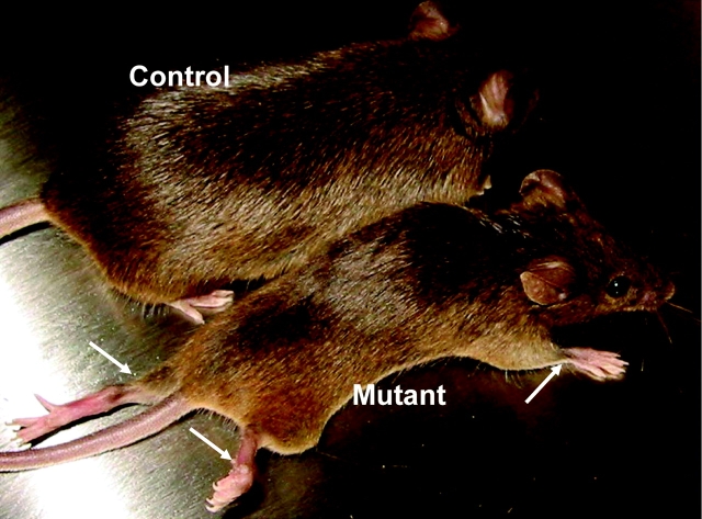
Laminins are heterotrimeric extracellular matrix proteins that regulate cell viability and function. Laminin-2, composed of alpha2, beta1, and gamma1 chains, is a major matrix component of the peripheral nervous system (PNS). To investigate the role of laminin in the PNS, we used the Cre-loxP system to disrupt the laminin gamma1 gene in Schwann cells. These mice have dramatically reduced expression of laminin gamma1 in Schwann cells, which results in a similar reduction in laminin alpha2 and beta1 chains. These mice exhibit motor defects which lead to hind leg paralysis and tremor. During development, Schwann cells that lack laminin gamma1 were present in peripheral nerves, and proliferated and underwent apoptosis similar to control mice. However, they were unable to differentiate and synthesize myelin proteins, and therefore unable to sort and myelinate axons. In mutant mice, after sciatic nerve crush, the axons showed impaired regeneration. These experiments demonstrate that laminin is an essential component for axon myelination and regeneration in the PNS.


Figures

Figure 1.
Figure 1.
Recombination of a floxed laminin γ1 allele by a CaMKIIα promoter-Cre transgene leads to reduced expression of laminin γ1 in the sciatic nerve. (A) Details of the laminin γ1 gene targeting construct. The two loxP sites (open triangles) were inserted into the laminin γ1 gene to flank exon 2 (E2). The neomycin resistance gene pGKneo (long open rectangle) used for ES cell selection was flanked by two frt sites (closed triangles). The P1 and P2 primer sites were used in B to monitor recombination by PCR. Restriction endonuclease sites are indicated (A, AvrII; H, HindIII; T, Tth111I; X, XcmI). (B) PCR analysis of tail DNA from wild-type and homozygous fLAMγ1 mice (f/f), and of DNA from various tissues from CaMKII/Cre:fLAMγ1 mice (f/f, CaMKII-Cre/+). The primers used allow amplification of the wild-type allele and the unrecombined and recombined fLAMγ1 allele. In tissues where recombination takes place, both unrecombined and recombined alleles are detected because there are some cells (e.g., vascular tissue) in which recombination does not occur. Recombination is prominent in the hippocampus (Hip) as expected, and also in the spinal cord (SC) and sciatic nerve (SN); but it is minimal in the skeletal muscle from the leg. (C) Transverse sections of sciatic nerve from control and mutant mice at P1 were double stained for laminin γ1 (a and c) and S-100 (b and d). (a) The nerve from a control animal shows strong laminin γ1 staining; (c) nerve from a mutant mouse shows greatly reduced expression of laminin γ1 (arrowheads) but there was still some expression (closed arrows). Laminin expression in the epineurium is not affected (open arrows). Even though laminin γ1 expression is dramatically decreased in mutant sciatic nerve, the S-100 staining was similar to control (b and d). Con, control; Mt, mutant.
Figure 2.
Figure 2.
Nerve axons do not express laminin γ1. E17.5 control embryo sections were stained for neurofilament (A) and laminin γ1 (B). The sciatic nerves were identified by the location of the neurofilament staining. At this developmental stage, some axonal bundles have not yet been sorted by Schwann cells (A and C, green), and these axons do not show laminin γ1 staining (B). However, the epineurium and surrounding tissues showed strong laminin γ1 staining (B and C, arrowheads).
Figure 3.
Figure 3.
Reduced expression of laminin α2, β1, and γ1 chains in the sciatic nerve of CaMKII/Cre:fLAMγ1 mice. Serial transverse sections of control (A–C) and mutant (D–F) mouse sciatic nerves at P28 were stained for laminin α2 (A and D), β1 (B and E), and γ1 (C and F). The expression of these laminin chains (components of laminin-2) were dramatically reduced in the mutant mice and showed similar staining patterns on adjacent sections (compare D, E, and F). Western blot analysis using the same monoclonal antibodies showed that expression of laminin α2 (G), β1 (H), and γ1 (I) in mutant sciatic nerves was dramatically reduced. Con, control sciatic nerve; Mt, mutant sciatic nerve.
Figure 4.
Figure 4.
CaMKII/Cre:fLAMγ1 mice have motor dysfunction. The mouse on the left was homozygous for the fLAMγ1 allele, but did not contain the CaMKII-Cre gene. Mice of this genotype or mice heterozygous for the fLAMγ1 allele and carrying the CaMKII-Cre gene were always normal. The mouse on the right (Mutant) was homozygous for the fLAMγ1 allele and carried the CaMKII-Cre gene. These mice were smaller and exhibited muscle weakness and in most cases complete paralysis of the legs (arrows) and muscular atrophy.
Figure 5.
Figure 5.
Nerves from CaMKII/Cre:fLAMγ1 and P0/Cre:fLAMγ1 mice have defective axonal sorting and myelination. (A–H) Transverse sections of sciatic nerve from control (A, C, E, and G) and CaMKII/Cre:fLAMγ1 mutant mice (B, D, F, and H) were examined at various ages by Richardson's staining. The age of the mouse is indicated on the left of each panel. In the controls, even though there were some unsorted axonal bundles in P1 (A, arrows), the axons were gradually subdivided and myelinated by Schwann cells (A, C, E, and G, arrowheads), and by P28 the myelination was complete (G). In contrast, in the mutant mice, the axonal subdivision and myelination was impaired and there were unsorted axon bundles (B, D, F, and H, arrows); and only a few axons became myelinated (D and F, arrowheads). By P28, there were large bundles of unsorted axons and few myelinated fibers (H, arrowhead). (I–L) Comparison of changes in ventral and dorsal spinal root in adult mutant mice. Transverse sections were stained with Richardson's staining. Both ventral (motor) and dorsal (sensory) roots were affected, but the ventral root appeared more severe. The roots and the nerve were similarly affected (H, J, L, and controls in I and K). Sciatic nerves from P0/Cre:fLAMγ1 mutant mice at P28 (N, control in M) showed a similar phenotype to those from CaMKII/Cre:fLAMγ1 mice. Arrows, unsorted axonal bundles; arrowheads, myelinated axons.
Figure 6.
Figure 6.
Schwann cells in CaMKII/Cre:fLAMγ1 nerves have normal proliferation and apoptosis during development. Longitudinal sciatic nerve sections of control (A) and mutant embryos (B) at E19.5 were stained for BrdU (red) and neurofilament (green) after a 1-h pulse of BrdU, and the images were merged. In C and D, the sciatic nerve sections at E19.5 were stained with TUNEL (red), for neurofilament (green) and counterstained with DAPI (blue), and the images were merged. Schwann cells in mutant mice had similar nuclei BrdU incorporation as controls, and populated the nerve (A and B, arrows). Statistical analysis using the Mann-Whitney U test revealed no significant difference in percentage of BrdU-incorporated nuclei between control and mutant embryos (E). TUNEL staining showed that the ratio of positive nuclei was similar in mutant and the controls at E17.5, E19.5, and P5 (E). The TUNEL-positive nuclei always overlapped with DAPI staining (C and D, arrows). Error bars represent the SEM.
Figure 7.
Figure 7.
Schwann cells that lack laminin γ1 are present in nerves but do not differentiate. Sections of sciatic nerve from P28 d mutant mice were stained for S-100 (A and G), laminin γ1 (B and H), Krox-20 (C), and MBP (I). The images in A–C are merged in D. After photography, the sections were counterstained with DAPI, which is much stronger than the previous laminin staining using the same fluorescence dye, and therefore only showed DAPI staining (E). The DAPI-stained images were merged with S-100 (A) and Krox-20 (C) stained images and shown in F. The staining showed an S-100–positive Schwann cell (A, arrow) that did not express laminin γ1 (B, arrow) and did not show nuclei Krox-20 staining (C–F, arrow). An S-100–positive Schwann cell with its nuclei shown on this section (A and E, arrowheads) did express laminin γ1 (B, arrowhead) and showed nuclear Krox-20 staining (C, D, and F, arrowhead). The nuclei of the rest of the Schwann cells not indicated in A were not on this section. The correlation of laminin γ1 expression and MBP synthesis was also compared. An S-100–positive Schwann cell (G, arrow) did not express laminin γ1 (H, arrow) and did not produce MBP (I and J, arrow), whereas the rest of the S-100–positive Schwann cells (G, arrowhead) expressed laminin γ1 and produced MBP (H–J, arrowhead). Bars, 5 μm.
Figure 8.
Figure 8.
Ultrastructure of CaMKII/Cre:fLAMγ1 sciatic nerve shows impairment of axon sorting and basal lamina formation. Electron microscopy of transverse sections of control (A) and mutant sciatic nerves (B) at P1. In control, many axonal bundles were sorted by Schwann cells, large axons have been segregated at the peripheral of the bundles (A, arrowheads) and many of them have formed a 1:1 relationship with a Schwann cell (A, arrows). In contrast, in the mutant nerve, the sorting was impaired, there were large axonal bundles (stars) but very few axons had been sorted (B, closed arrowhead) or formed a 1:1 relationship with a Schwann cell (B, arrows). Note that there were many Schwann cells in the mutant sciatic nerve (B, open arrowheads). At P15 in the mutant nerve, the unsorted axonal bundles contained different sized axons (C, stars). The Schwann cell closely associated with the naked axonal bundles (S2) sent abnormally thick processes between axons (arrows) but did not make myelin. Immediately adjacent to S2 is a Schwann cell (S1) with normal myelin. (D) Higher magnification of the boxed region in C shows that the myelinating Schwann cell S1 had a continuous basal lamina (arrowheads) attached to the cell membrane, whereas Schwann cell S2 did not form a continuous basal lamina (denuded areas indicated by thin arrows) and only had small patches of ECM attached to the cell membrane (thick arrow). Bars: (A and B) 2 μm; (C) 1 μm; (D) 0.5 μm.
Figure 9.
Figure 9.
Mutant sciatic nerves with reduced laminin γ1 expression are defective in axonal regeneration after injury. 1 or 28 d after sciatic nerve crush, the nerves were cut 7 mm distal to the lesion, and a crystal of the retrograde tracer fluororuby was applied to the cut nerve. On the uncrushed side, the sciatic nerve was cut at a similar place as the crushed side and a crystal of fluororuby was applied. 3 d later the spinal cords were analyzed for labeled motoneurons. (A) 1 d after crush, there were no labeled motoneurons on the crushed side but many on the uncrushed side, indicating that the crush was complete and there was no leakage of the dye. (B) 28 d after crush in the wild-type animal, most of the motoneurons on the crushed side were labeled (compare with the uncrushed side). (C) In contrast, in the mutant mice, the crushed side showed many fewer labeled motoneurons compared with the uncrushed side. (D) The percentage of labeled motoneurons in the crushed side compared with the uncrushed side in six control and six mutant mice was quantitated, and the difference between control and mutant animals was significant.
Figure 10.
Figure 10.
Model of laminin function in peripheral nerve myelination. During peripheral nerve development, laminin expressed in Schwann cell binds to integrin or other receptors. This may activate signaling pathways such as NF-κB p65/p50, allow proper axon sorting by Schwann cells, and activate genes for differentiation and myelination such as SCIP and Krox-20.

Similar articles

Cited by

References

    1. Akassoglou, K., K.W. Kombrinck, J.L. Degen, and S. Strickland. 2000. Tissue plasminogen activator-mediated fibrinolysis protects against axonal degeneration and demyelination after sciatic nerve injury. J. Cell Biol. 149:1157–1166. - PMC - PubMed
    1. Baron-Van Evercooren, A., A. Gansmuller, M. Gumpel, N. Baumann, and H.K. Kleinman. 1986. Schwann cell differentiation in vitro: extracellular matrix deposition and interaction. Dev. Neurosci. 8:182–196. - PubMed
    1. Bradley, W.G., and M. Jenkison. 1973. Abnormalities of peripheral nerves in murine muscular dystrophy. J. Neurol. Sci. 18:227–247. - PubMed
    1. Bunge, M.B. 1993. Schwann cell regulation of extracellular matrix biosynthesis and assembly. Peripheral Neuropathy. P.J. Dyck, P.K. Thomas, J. Griffin, P.A. Low, J.F. Poduslos, editors. W.B. Saunders, Philadelphia. 299–316.
    1. Bunge, R.P. 1993. Expanding roles for the Schwann cell: ensheathment, myelination, trophism and regeneration. Curr. Opin. Neurobiol. 3:805–809. - PubMed

Publication types

MeSH terms