. 2003 Nov 24;163(4):901-10.
doi: 10.1083/jcb.200304161.

Loss of proteolytically processed filaggrin caused by epidermal deletion of Matriptase/MT-SP1

Affiliations

Loss of proteolytically processed filaggrin caused by epidermal deletion of Matriptase/MT-SP1

Karin List et al. J Cell Biol. .

Abstract

Profilaggrin is a large epidermal polyprotein that is proteolytically processed during keratinocyte differentiation to release multiple filaggrin monomer units as well as a calcium-binding regulatory NH2-terminal filaggrin S-100 protein. We show that epidermal deficiency of the transmembrane serine protease Matriptase/MT-SP1 perturbs lipid matrix formation, cornified envelope morphogenesis, and stratum corneum desquamation. Surprisingly, proteomic analysis of Matriptase/MT-SP1-deficient epidermis revealed the selective loss of both proteolytically processed filaggrin monomer units and the NH2-terminal filaggrin S-100 regulatory protein. This was associated with a profound accumulation of profilaggrin and aberrant profilaggrin-processing products in the stratum corneum. The data identify keratinocyte Matriptase/MT-SP1 as an essential component of the profilaggrin-processing pathway and a key regulator of terminal epidermal differentiation.


Figures
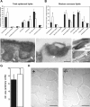
Figure 1.
Figure 1.
Matriptase/MT-SP1–deficient mice are born with epidermal lipid matrix defects and enlarged CEs. Total epidermal (A) and stratum corneum (B) lipids in Matriptase/MT-SP1−/− (open bars, n = 3) and control (solid bars, n = 3) neonates (mean weight percentage of total lipids ± SD). (A) *, P < 0.04 and (B) *, P < 0.02 (two-tailed t test). (C–F) Ultrastructural appearance of transitional cells (C and D) and stratum corneum (E and F) of control epidermis (C and E) and Matriptase/MT-SP1−/− epidermis (D and F) of ruthenium-stained sections. (C) Lipid lamellar bodies with well-aligned lipid stacks (arrows) at the interface between the transitional layer and stratum corneum in control epidermis. (D) Rare and poorly formed lamellar bodies (arrow) with wavy, short, and disorganized lipid structures in Matriptase/MT-SP1−/− epidermis. (E) Highly ordered intercorneocyte lipid lamellar membranes (arrow) in control stratum corneum. (F) Poorly formed intercorneocyte lipid lamellae in Matriptase/MT-SP1−/− stratum corneum (arrow). (G) Average surface area of newborn Matriptase/MT-SP1−/− (open bar, n = 70) and littermate control (solid bar; n = 50) CEs (mean ± SD; *, P < 0.004; two-tailed t test). The experiment was repeated three times with similar results. (H) Morphological appearance of purified CEs from Matriptase/MT-SP1−/− epidermis (right panel) and littermate control epidermis (left panel) visualized by phase-contrast light microscopy. Bars: (C–F) 100 nm, (H) 30 μm.
Figure 2.
Figure 2.
Impaired desquamation of newborn Matriptase/MT-SP1–deficient stratum corneum. (A) Macroscopic appearance of neonatal dorsal skin of control (left) and littermate Matriptase/MT-SP1−/− (right) after 12 sequential applications of adhesive-coated discs. The stratum corneum is completely removed from the tape-stripped area of the control epidermis, exposing the lower epidermal layers, whereas only minimal defects are apparent in Matriptase/MT-SP1−/− epidermis subjected to the same treatment. Arrowheads indicate the outline of the tape-stripped area. (B) Microscopic appearance of control (left panels) and littermate Matriptase/MT-SP1−/− (right panels) epidermis before (top panels) and after (bottom panels) 12 rounds of tape stripping. Complete removal of the stratum corneum with exposed upper granular and transitional cell layers in control mice and minimal stratum corneum removal in Matriptase/MT-SP1−/− epidermis. Hematoxylin and eosin staining. “E” indicates the location of basal and suprabasal epidermis, and “S” indicates the location of the stratum corneum. Bars, 20 μm. (C) Accumulated removal of extractable stratum corneum proteins from Matriptase/MT-SP1−/− (open squares, n = 7) and littermate control (closed circles, n = 7) mice after tape stripping (± SEM; *, P < 0.03; two-tailed t test).
Figure 3.
Figure 3.
Matriptase/MT-SP1–deficient epidermis lacks proteolytically processed filaggrin monomer. (A) Amido black staining (lanes 1–4) of SDS-PAGE–separated protein extracts from epidermis (lanes 1 and 2) and dermis (lanes 3 and 4), and whole-skin extract filaggrin monomer Western blot (lanes 5 and 6) of control (lanes 1, 3, and 5) and littermate Matriptase/MT-SP1−/− (lanes 2, 4, and 6) neonates. (B) Western blot analysis of SDS-PAGE–separated proteins from skin from control (lane 1) and littermate Matriptase/MT-SP1−/− (lane 2) neonates using antibodies against the basal layer markers keratin-5 and -14, the suprabasal layer markers keratin-1 and -10, and the granular layer/stratum corneum marker loricrin. (C) Western blot analysis of total skin protein extracts from neonates (lanes 1 and 2) and E18.5 embryos (lanes 3 and 4) from control (lanes 1 and 3) and littermate Matriptase/MT-SP1−/− (lanes 2 and 4) mice using the filaggrin monomer repeat antibody. (D) Western blot of total skin protein extracts from newborn control (lane 1), littermate Matriptase/MT-SP1−/− (lane 2), Klf4 control (lane 3), and littermate Klf4−/− (lane 4) neonates using the filaggrin monomer repeat antibody. Arrowheads in A, C, and D indicate the 32-kD filaggrin monomer immunoreactive protein specifically missing in Matriptase/MT-SP1−/− epidermis. The positions of mol wt markers (kD) are indicated.
Figure 4.
Figure 4.
Defective proteolytic processing of profilaggrin and incorporation of aberrantly processed profilaggrin into the stratum corneum of Matriptase/MT-SP1–deficient epidermis. (A) Profilaggrin (Pro-FG) and profilaggrin-derived (FG) proteins were partially purified by NaSCN and urea extraction of the skin of control (lanes 1 and 3) and littermate Matriptase/MT-SP1−/− (lanes 2 and 4) neonates. 50 (lanes 1 and 2) or 0.5 (lanes 3 and 4) μg protein was separated by SDS-PAGE under reducing conditions and stained with Coomassie brilliant blue (lanes 1 and 2) or subjected to Western blot analysis with a filaggrin monomer antibody (lanes 3 and 4). The positions of profilaggrin-related protein products are indicated on the left, and the positions of mol wt markers (kD) on the right. (B–E) Representative examples of filaggrin immuno-EM of filaggrin granules (F) in granular layer cells (B and C), and corneocytes (“c” in D and E) from littermate control (B and D) and Matriptase/MT-SP1−/− (C and E) epidermis. Filaggrin antibody binding was visualized with colloidal gold-labeled secondary antibodies. Both filaggrin granules in granular layer cells (B and C) and keratin aggregates of terminally differentiated corneocytes (D and E, arrowheads) display intense gold labeling, indicative of incorporation of profilaggrin immunoreactive material, with no detectable differences in the labeling intensity in control and Matriptase/MT-SP1−/− epidermis. Bars: (B and C) 100 nm, (D and E) 75 nm.
Figure 5.
Figure 5.
Loss of proteolytic liberation of NH 2 -terminal filaggrin S-100–containing protein products in Matriptase/MT-SP1–deficient epidermis. Western blot analysis of epidermal protein extracts from control (lane 1) and littermate Matriptase/MT-SP1−/− (lane 2) neonates stained with the filaggrin S-100 A domain antibody. Control epidermis contains mature S-100 protein (28- and 30-kD doublets) and the isolated 16-kD S-100 A domain. In contrast, Matriptase/MT-SP1−/− epidermis contains no detectable S-100 protein, a dramatic accumulation of the 50-kD processing intermediate, and accumulation of an unidentified 10-kD filaggrin-related protein product. “A” and “B” indicate the A and B domains of the filaggrin S-100 protein, “TGF” indicates the truncated filaggrin repeat, and “FG 1” indicates the first filaggrin repeat.
Figure 6.
Figure 6.
Prolonged exposure of transplanted Matriptase/MT-SP1 epidermis to air leads to severe hyperproliferative ichthyosis. Gross (A) and microscopic (B–G) appearance of control (B, D, and F) and Matriptase/MT-SP1−/− (C, E, and G) skin, 3 wk after transplantation to nude mice. Sections were stained with hematoxylin and eosin (B and C), or by immunostaining with anti-BrdU (D and E) or keratin-6 (F and G) antibodies. (A) Alopecia, grotesque epidermal thickening, and formation of epidermal plates in transplanted Matriptase/MT-SP1−/− skin. (B–G) Profound acanthosis and hyperkeratosis of transplanted Matriptase/MT-SP1−/− skin (compare B with C) with epidermal hyperproliferation (compare D with E) and keratin-6 overexpression (compare F with G). “E” in B and C indicates the location of basal and suprabasal epidermis, and “S” indicates the location of the stratum corneum. Examples of proliferating keratinocytes in the hair follicles (arrows) in D and in the epidermis in E are indicated with arrowheads. Keratin-6 staining of hair follicles (but not epidermis) in F, compared with strong keratin-6 staining of both epidermis and hair follicles in G. Bars, (B–G) 20 μm.

Similar articles

Cited by

References

    1. Akiyama, M. 1999. The pathogenesis of severe congenital ichthyosis of the neonate. J. Dermatol. Sci. 21:96–104. - PubMed
    1. Compton, J.G., J.J. DiGiovanna, K.A. Johnston, P. Fleckman, and S.J. Bale. 2002. Mapping of the associated phenotype of an absent granular layer in ichthyosis vulgaris to the epidermal differentiation complex on chromosome 1. Exp. Dermatol. 11:518–526. - PubMed
    1. Dale, B.A., K.A. Holbrook, and P.M. Steinert. 1978. Assembly of stratum corneum basic protein and keratin filaments in macrofibrils. Nature. 276:729–731. - PubMed
    1. Dale, B.A., K.A. Holbrook, J.R. Kimball, M. Hoff, and T.T. Sun. 1985. Expression of epidermal keratins and filaggrin during human fetal skin development. J. Cell Biol. 101:1257–1269. - PMC - PubMed
    1. Dale, B.A., K.A. Holbrook, P. Fleckman, J.R. Kimball, S. Brumbaugh, and V.P. Sybert. 1990. Heterogeneity in harlequin ichthyosis, an inborn error of epidermal keratinization: variable morphology and structural protein expression and a defect in lamellar granules. J. Invest. Dermatol. 94:6–18. - PubMed

Publication types

MeSH terms