Comparative Study
. 2003 Dec;122(6):713-26.
doi: 10.1085/jgp.200308891.

Interactions between electron and proton currents in excised patches from human eosinophils

Affiliations
Comparative Study

Interactions between electron and proton currents in excised patches from human eosinophils

Gabor L Petheö et al. J Gen Physiol. 2003 Dec.

Abstract

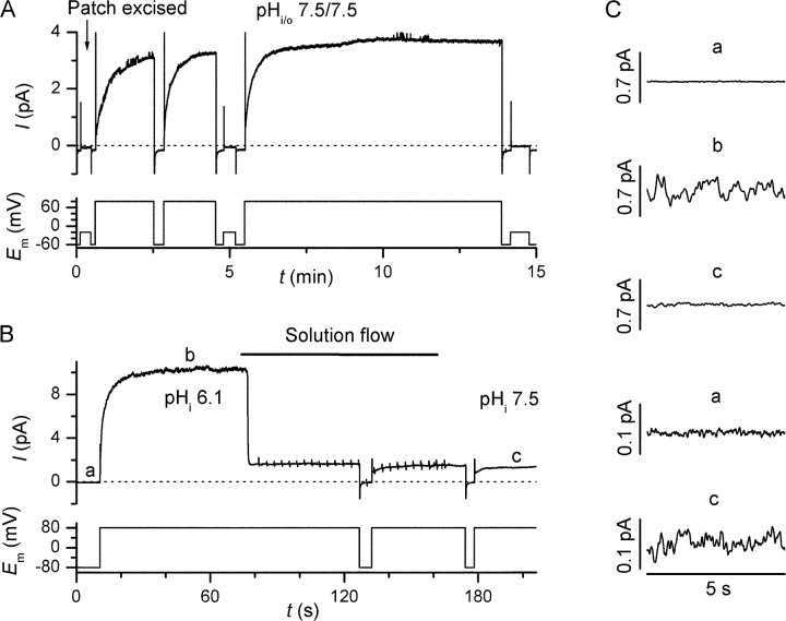
The NADPH-oxidase is a plasma membrane enzyme complex that enables phagocytes to generate superoxide in order to kill invading pathogens, a critical step in the host defense against infections. The oxidase transfers electrons from cytosolic NADPH to extracellular oxygen, a process that requires concomitant H+ extrusion through depolarization-activated H+ channels. Whether H+ fluxes are mediated by the oxidase itself is controversial, but there is a general agreement that the oxidase and H+ channel are intimately connected. Oxidase activation evokes profound changes in whole-cell H+ current (IH), causing an approximately -40-mV shift in the activation threshold that leads to the appearance of inward IH. To further explore the relationship between the oxidase and proton channel, we performed voltage-clamp experiments on inside-out patches from both resting and phorbol-12-myristate-13-acetate (PMA)-activated human eosinophils. Proton currents from resting cells displayed slow voltage-dependent activation, long-term stability, and were blocked by micromolar internal [Zn2+]. IH from PMA-treated cells activated faster and at lower voltages, enabling sustained H+ influx, but ran down within minutes, regaining the current properties of nonactivated cells. Bath application of NADPH to patches excised from PMA-treated cells evoked electron currents (Ie), which also ran down within minutes and were blocked by diphenylene iodonium (DPI). Run-down of both IH and Ie was delayed, and sometimes prevented, by cytosolic ATP and GTP-gamma-S. A good correlation was observed between the amplitude of Ie and both inward and outward IH when a stable driving force for e- was imposed. Combined application of NADPH and DPI reduced the inward IH amplitude, even in the absence of concomitant oxidase activity. The strict correlation between Ie and IH amplitudes and the sensitivity of IH to oxidase-specific agents suggest that the proton channel is either part of the oxidase complex or linked by a membrane-limited mediator.


Figures

F<sc>igure</sc> 1.
Figure 1.
Depolarization-activated, pHi-sensitive currents in inside-out patches from nonstimulated human eosinophils. Human eosinophils were adhered to glass coverslips in the absence of soluble stimuli and patched within 10 min of plating. (A) After patch excision at −20 mV (arrow) depolarization to 80 mV evoked time-dependent currents that activated slowly and were stable for several minutes. The voltage protocol is displayed below the current traces. (B) Effect of bath alkalization from pH 6.1 to 7.5 on the current amplitude at 80 mV. Spikes on the current trace during solution change are capacitance artifacts caused by the suction pump. Trace is representative from six experiments with similar results. (C) Excess fluctuations associated with the outward current (b and c), are compared with the background noise measured at ∼E H (a). Traces a and c are shown of two different scales to match the steady-state current amplitude measured at pHi 6.1. Time bar applies to all traces in C.
F<sc>igure</sc> 2.
Figure 2.
Effect of pHi change on E rev. To determine the reversal potential (E rev) of the depolarization-activated current, we took advantage of its slow activation kinetic. Every 25 s, a long-lasting (15 s, a) or short-lasting (1 s, b) activating pulse to 80 mV was applied, followed by a rapid voltage ramp to the −100-mV holding potential (C). This voltage protocol was applied at different transmembrane pH gradients (A and B), and the reversal potential of the voltage- and time-dependent currents (D) was determined by subtracting the two “ramp tail” currents (a − b). Increasing the pHi from 6.1 to 7.5 caused an ∼75-mV shift in the 0 current potential (E rev). Traces are low-pass filtered at 30 Hz and are representatives of six experiments with similar results.
F<sc>igure</sc> 3.
Figure 3.
Effects of intracellular Zn2+ and Ca2+ on the proton currents. Voltage steps were applied every 15 s from the −80 mV holding potential to −70 mV and then to test potentials ranging from −60 to 80 mV in 20-mV increments. Current traces were acquired under control conditions (A and D) or in the presence of 45 μM free Zn2+ (B) or 8 μM Ca2+ (E). Traces were low-pass filtered at 20 Hz, pHi/o was 7.43–7.47/7.5, citrate concentration in the bath was 3 mM in all cases. (C and F) Effect of 45 μM Zn2+ (n = 5, •, control; ▿, Zn2+) and 8 μM Ca2+ (n = 4, ▪, control; ▵, Ca2+) on the near steady-state current-voltage relationship. The average current during the last second of the 8-s long voltage step was measured at different test potentials. The measured current amplitudes at each test potential were normalized to the control value measured at 80 mV in the same patch. Time scale applies to all current traces. Traces in A and B and D and E have the same current scale.
F<sc>igure</sc> 4.
Figure 4.
Proton current in patches excised from PMA-stimulated eosinophils. Human eosinophils were adhered to glass coverslips in the presence of 200–400 nM PMA and patched after 5–10 min of stimulation. (A) Progressive run-down of H+ current could be observed when patches were excised (arrow) into ATP and GTP-γ-S–free bath solution. Stepwise changes with unexpectedly large amplitude (insert) were present in the course of run-down in 14 of 16 patches excised into nucleotide-free solution. Note the break in the time axis. (B) Absence of run-down in a patch excised in a bath solution containing 3 mM ATP and 20 μM GTP-γ-S. When indicated, the bath was exchanged to a nucleotide-free solution. The increase in H+ current amplitude during solution flow probably reflects the mechanosensitivity of H+ channels in the excised patch (unpublished data). Trace is representative of six experiments with similar results. In one case stepwise changes were observed after washing out the nucleotides. (C) Currents from B elicited by a pulse to 80 mV were normalized to the steady-state current amplitude to demonstrate the difference between the activation kinetics before (a) and after (b) run-down.
F<sc>igure</sc> 5.
Figure 5.
Properties and run-down of inward and outward H+ currents. Patches excised from PMA stimulated eosinophils were recorded in a bath solution containing 5 mM ATP and 25 μM GTP-γ-S. Inward and outward H+ currents were elicited by pulses to 0 and 60 mV, the driving forces for H+ current at these voltages were approximately −30 and 30 mV, respectively. In every patch exhibiting run-down, the two currents decreased in parallel (n > 20). Note the break in the time axis. Inset demonstrates excess low-frequency fluctuations associated with the inward H+ current recorded at 0 mV (a) as compared with the background noise recorded at −60 mV (b). Time and current bars apply for both traces in the insert.
F<sc>igure</sc> 6.
Figure 6.
Electron currents and superoxide production in patches excised from PMA-treated eosinophils. (A) Bath application of 4 mM NADPH (arrowhead) evoked an electron current at −60 mV that was blocked by 2 μM DPI (arrow). (B) A patch excised with a pipette containing 0.5 mg/ml NBT was photographed shortly before (left) and 15 min after (right) bath application of 0.8 mM NADPH. Note the formation of a dark NBT precipitate (formazan) due to O2 .− production near the pipette tip. The patch was excised from the cell at 11 o'clock from the pipette tip. (C) The e current evoked by 0.8 mM NADPH (arrow) in the presence of 0.5 mg/ml pipette NBT faded out with time in a stepwise manner. Traces are representatives of the experiments used to establish I eI H plots (Fig. 7), in which the amplitude of the inward H+ current was measured at 0 mV just before stepping back to −60 mV (a), whereas the e current was measured as the difference between the holding current amplitude at −60 mV in the absence and presence of NADPH (b and c).
F<sc>igure</sc> 7.
Figure 7.
Correlation between inward H+ and e currents. Proton and electron currents were evoked and measured as described in Fig. 6. Electron currents were induced either by bath application of 4 mM (A) or 0.8 mM NADPH in the absence (B) or presence (C) of 0.5 mg/ml NBT in the pipette solution. Steady-state (A and B) or peak (C) e current amplitudes at −60 mV are plotted against the average inward H+ current amplitude during the last 5 s at 0 mV. Dotted lines are the results of linear regressions constrained to pass through the origin (R > 0.91 for panel A and R > 0.98 for panel C). Solid line in C is the result of fitting a first order decaying exponential curve to the data (R 2 > 0.98) to demonstrate apparent saturation of electron transport, probably reflecting the limitation in electron current amplitude due to NBT precipitation.
F<sc>igure</sc> 8.
Figure 8.
Effects of DPI and NADPH on the proton current. (A) Effect of sequential bath application of 2 μM DPI (arrowhead) and 0.8 mM NADPH (arrow) on inward and outward H+ currents. Note the transient increase in the inward current amplitude preceding the inhibition of the H+ current after the application of NADPH. The patch was excised from a PMA-treated cell. The bath solution contained 5 mM ATP and 25 μM GTP-γ-S. (B) Summary of the effect of 2 μM DPI and 0.8 mM NADPH on the inward H+ current with NADPH or DPI added first (left and right, respectively, n = 5 each). Single asterisks denote significant difference from control values, double asterisk indicates significant difference between columns. (C) Summary of the combined effect of 2 μM DPI and 0.8 mM NADPH on the inward and outward H+ current (n = 7). Data are compiled from Fig. 8 B, irrespective of the sequence of addition. Asterisk indicates significant difference from control values. To remove the potential contribution of e currents, a biexponential fit was applied to the depolarization-activated H+ currents, extrapolated to the zero and steady-state values (for the 60-mV pulse only), and the difference taken as H+ current amplitude.

Similar articles

Cited by

References

    1. Babior, B.M., J.D. Lambeth, and W. Nauseef. 2002. The neutrophil NADPH oxidase. Arch. Biochem. Biophys. 397:342–344. - PubMed
    1. Banfi, B., A. Maturana, S. Jaconi, S. Arnaudeau, T. Laforge, B. Sinha, E. Ligeti, N. Demaurex, and K.H. Krause. 2000. A mammalian H+ channel generated through alternative splicing of the NADPH oxidase homolog NOH-1. Science. 287:138–142. - PubMed
    1. Banfi, B., G. Molnar, A. Maturana, K. Steger, B. Hegedus, N. Demaurex, and K.H. Krause. 2001. A Ca2+-activated NADPH oxidase in testis, spleen, and lymph nodes. J. Biol. Chem. 276:37594–37601. - PubMed
    1. Banfi, B., J. Schrenzel, O. Nusse, D.P. Lew, E. Ligeti, K.H. Krause, and N. Demaurex. 1999. A novel H+ conductance in eosinophils: unique characteristics and absence in chronic granulomatous disease. J. Exp. Med. 190:183–194. - PMC - PubMed
    1. Biberstine-Kinkade, K.J., F.R. DeLeo, R.I. Epstein, B.A. LeRoy, W.M. Nauseef, and M.C. Dinauer. 2001. Heme-ligating histidines in flavocytochrome b(558): identification of specific histidines in gp91(phox). J. Biol. Chem. 276:31105–31112. - PubMed

Publication types