Comparative Study
. 2003 Dec;122(6):795-804.
doi: 10.1085/jgp.200308953.

Ligand-induced closure of inward rectifier Kir6.2 channels traps spermine in the pore

Affiliations
Comparative Study

Ligand-induced closure of inward rectifier Kir6.2 channels traps spermine in the pore

L Revell Phillips et al. J Gen Physiol. 2003 Dec.

Abstract

Small organic amines block open voltage-gated K+ channels and can be trapped by subsequent closure. Such studies provide strong evidence for voltage gating occurring at the intracellular end of the channel. We engineered the necessary properties (long block times with unblock kinetics comparable to, or slower than, the kinetics of gating) into spermine-blocked, ATP-gated (N160D,L157C) mutant KATP channels, in order to test the possibility of "blocker trapping" in ligand-gated Kir channels. Spermine block of these channels is very strongly voltage dependent, such that, at positive voltages, the off-rate of spermine is very low. A brief pulse to negative voltages rapidly relieves the block, but no such relief is observed in ATP-closed channels. The results are well fit by a simple kinetic model that assumes no spermine exit from closed channels. The results incontrovertibly demonstrate that spermine is trapped in channels that are closed by ATP, and implicate the M2 helix bundle crossing, or somewhere lower, as the probable location of the gate.


Figures

F<sc>igure</sc> 1.
Figure 1.
Location of possible gates in Kir6.2 channels. (A) Cartoon showing a likely pore structure for Kir6.2 (model based on KcsA; Loussouarn et al., 2000). Backbone locations of critical residues mutated in the present study are indicated. Also shown is a space-filling model of the elongated form of spermine, placed in a likely blocking location in the pore axis. (B) Models for channel gating. In each case, spermine has voltage-dependent access to its binding site in the open state, but in the ATP-closed state may still access the binding site if gating occurs at the selectivity filter (Scheme I), or has no access if gating occurs at, or more cytoplasmically to, the M2 helix bundle crossing (Scheme II).
F<sc>igure</sc> 2.
Figure 2.
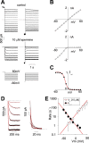
Voltage-dependent block of N160D/L157C channels by spermine. (A) Representative inside-out current records in the absence and presence of 10 μM spermine, in response to voltage steps from Vh = −60 mV to voltages between −60 and +60 mV (10-mV steps). (B) Steady-state current-voltage relationships (from A). (C) Relative conductance (Irel)-voltage relationships computed from above (Irel = Ispermine/Icont), fit with Boltzmann function (Z = 4.04, V1/2 = −14.4 mV). (D) Expanded time base records of current in the presence of spermine, in response to voltage steps from +60 mV to voltages between −10 and −60 mV (left), or from −60 mV to voltages between +10 and +60 mV (right), with single exponential fits indicated. (E) Spermine entry (kin × [spermine]) and exit (kout) rates as a function of Vm, estimated from current relaxation kinetics (D) and steady-state block (C) by 10 μM spermine (see text).
F<sc>igure</sc> 3.
Figure 3.
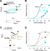
Spermine unblock rates estimated using “concentration jump.” (A) Spermine (10 μM) was briefly applied (as indicated) to block all channels at +40 mV. After spermine removal, currents recover slowly (τ ∼20 s). Brief pulses to voltages more negative than −20 (left and expanded) significantly relieve inhibition. (B) Relative recovery of current is plotted versus pulse voltage. The solid red line indicates the predicted recovery given the spermine off-rate (kout) calculated in Fig. 2. (C) Spermine (10 μM) was briefly applied (as indicated) to block all channels at +40 mV. After spermine removal, pulses to −40 mV were applied for various durations. (D) Relative recovery of current is plotted versus pulse duration for four similar experiments (mean ± SEM). The solid red line indicates the predicted recovery given the spermine off-rate calculated in Fig. 2.
F<sc>igure</sc> 4.
Figure 4.
ATP closure traps spermine in the channel. (A) Spermine (10 μM) was briefly applied (as indicated) to block N160D/L157C channels at +40 mV. After spermine removal, currents recover slowly (trace 1). In trace 2, after spermine removal, a 300-ms pulse to −40 mV completely relieves block. In trace 3, channels were exposed to 10 mM ATP after spermine removal. A 300-ms pulse to −40 mV was applied, then ATP removed. In the ATP closed channels, there is no recovery from spermine block. (B) In trace 2, spermine (10 μM) was briefly applied (as indicated) to block Kir6.2[L164C, N160D] channels at +40 mV. After spermine removal, a 100-ms pulse to −40 mV completely relieves block. In trace 3, channels were exposed to 10 mM ATP following spermine removal. In contrast to N160D/L157C channels, ATP fails to close N160D/L164C channels, and a 100-ms pulse to −40 mV applied during ATP exposure fails to protect against spermine release (representative experiment, n = 3).
F<sc>igure</sc> 5.
Figure 5.
ATP traps spermine in closed channels. (A) Spermine (10 μM) was briefly applied to block N160D/L157C channels at +40 mV, and then spermine was removed ∼2 s before the onset of the recorded traces and the patch was exposed to 10 mM ATP (rPo = 0.10) at the onset of each recording. Variable duration pulses to −40 mV were applied. ATP was subsequently removed, and the extent of recovery assessed as indicated (current was averaged for 1 s at dashed vertical line). Shown in gray is the response to a 100-ms pulse to −40 mV without exposure to ATP (rPo = 1.0). This completely relieves block in open channels. (B) Relative recovery of current is plotted versus pulse duration (from the experiment in A). The solid lines indicate the predicted recovery for Po = 1.0 and Po = 0.1 for Schemes I and II (see text). (C) Spermine (10 μM) was briefly applied to block N160D/L157C channels at +40 mV, and then spermine was removed ∼2 s before the onset of the recorded traces. A 1-s pulse to −40 mV completely relieves block of open channels (rPo = 1.0, black trace). The protocol was repeated and, after removal of spermine, the patch was exposed to 10 mM ATP (blue trace, rPo ∼0.1) at the beginning of the recording. A 1-s pulse to −40 mV was applied and recovery assessed as indicated. The patch was then exposed to 5 mg/ml PIP2 for ∼1 min to increase channel open state stability. Exposure to PIP2 increased rPo in 10 mM ATP to ∼0.5. The patch was again blocked by spermine (10 μM) and, after spermine removal, the patch was exposed to 10 mM ATP (orange trace, rPo = 0.50), and a 1-s step to −40 mV was applied and recovery assessed as indicated. (D) Relative recovery of current is plotted versus pulse duration. The solid lines indicate the predicted recovery for Po for Scheme II (see text). The trapping effect of ATP is antagonized by PIP2.
SCHEME I
SCHEME I
SCHEME II
SCHEME II
F<sc>igure</sc> 6.
Figure 6.
Spermine trapping depends on open probability. Relative recovery of current is plotted versus pulse duration for multiple similar experiments of the kind shown in Fig. 5. Data from experiments (as in Fig. 5, A and C) were pooled by rPo, without regard to applied [ATP] and without regard to prior exposure to PIP2. The solid lines indicate the predicted recovery for Po as indicated from Scheme II (see text). The pooled data correspond to rPo = 1, n = 12 (black symbols); rPo = 0.49 ± 0.06, n = 4 (orange symbols); rPo = 0.09 ± 0.01, n = 5 (blue symbols); rPo = 0.013 ± 0.005, n = 3 (purple symbols).
SCHEME III
SCHEME III

References

    1. Armstrong, C.M. 1966. Time Course of TEA(+)-induced Anomalous Rectification in Squid Giant Axons. J. Gen. Physiol. 50:491–503. - PMC - PubMed
    1. Armstrong, C.M. 1969. Inactivation of the potassium conductance and related phenomena caused by quaternary ammonium ion injection in squid axons. J. Gen. Physiol. 54:553–575. - PMC - PubMed
    1. Armstrong, C.M. 1971. Interaction of tetraethylammonium ion derivatives with the potassium channels of giant axons. J. Gen. Physiol. 58:413–437. - PMC - PubMed
    1. Baukrowitz, T., U. Schulte, D. Oliver, S. Herlitze, T. Krauter, S.J. Tucker, J.P. Ruppersberg, and B. Fakler. 1998. PIP2 and PIP as determinants for ATP inhibition of KATP channels. Science. 282:1141–1144. - PubMed
    1. Bruening-Wright, A., M.A. Schumacher, J.P. Adelman, and J. Maylie. 2002. Localization of the activation gate for small conductance Ca2+-activated K+ channels. J. Neurosci. 22:6499–6506. - PMC - PubMed

Publication types

MeSH terms