Review
. 2003 Dec 9;42(48):14083-91.
doi: 10.1021/bi035656u.

Mechanism of aromatic amino acid hydroxylation

Affiliations
Review

Mechanism of aromatic amino acid hydroxylation

Paul F Fitzpatrick. Biochemistry. .
No abstract available


Figures

Scheme 1
Scheme 1
Scheme 2
Scheme 2
Scheme 3
Scheme 3
Scheme 4
Scheme 4
Scheme 5
Scheme 5
Scheme 6
Scheme 6
Figure 1
Figure 1
(A) Overlay of the catalytic domains of human PheH (PDB file 1J8U), rat TyrH (PDB file 2TOH), human TrpH (PDB file 1MLW), and C. violaceum PheH (PDB file 1LTV) (B) Combined catalytic and regulatory domains of rat PheH. The structure is based on the PDB file 1PHZ; the protein used for structure determination lacked the C-terminal 24 residues, and the N-terminal 18 residues are not seen in the structure. The orientation is the same as in panel A. (C) Model for the intact rat PheH tetramer. The model was constructed by superimposing the catalytic and regulatory domains of rat PheH shown in panel B with the structure of the tetrameric TyrH catalytic domain (PDB file 2TOH).
Figure 2
Figure 2
Comparison of the iron and tetrahydrobiopterin binding sites of PheH in the absence (A) and presence (B) of β-thienylalanine. The structures are from the PDB files 1J8U and 1KWO.
Figure 3
Figure 3
Amino acid substrate binding site of PheH. The structure is from the PDB file 1KWO.
Figure 4
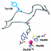
Figure 4
Movement of the 130–150 loop in PheH upon binding of β-thienylalanine to the tetrahydrobiopterin bound enzyme. The loop with Tyr138 in blue is from the binary complex (PDB file 1J8U); the positions of the active site iron, the substrates, and the metal ligands are from the ternary complex (PDB file 1KWO).

Similar articles

Cited by

References

    1. Eisensmith RC, Woo SLC. Mol. Biol. Med. 1991;8:3–18. - PubMed
    1. Ledley FD, DiLella AG, Kwok SCM, Woo SLC. Biochemistry. 1985;24:3389–3394. - PubMed
    1. Grenett HE, Ledley FD, Reed LL, Woo SLC. Proc. Natl. Acad. Sci. U.S.A. 1987;84:5530–5534. - PMC - PubMed
    1. Daubner SC, Lohse DL, Fitzpatrick PF. Protein Sci. 1993;2:1452–1460. - PMC - PubMed
    1. Daubner SC, Hillas PJ, Fitzpatrick PF. Biochemistry. 1997;36:11574–11582. - PubMed

Publication types

LinkOut - more resources