Acute exposure of ceramide enhances cardiac contractile function in isolated ventricular myocytes
- PMID: 14645138
- PMCID: PMC1574130
- DOI: 10.1038/sj.bjp.0705510
Acute exposure of ceramide enhances cardiac contractile function in isolated ventricular myocytes
Abstract
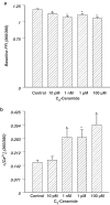
1. The sphingolipid ceramide, a primary building block for all other sphingolipids, is associated with growth arrest, apoptosis, and lipotoxic dysfunction. Interestingly, ceramide may attenuate high glucose-induced myocyte dysfunction, produce Ca2+ influx, and augment smooth muscle contraction. To determine the role of ceramide on cardiac excitation-contraction (E-C) coupling, electrically paced adult rat ventricular myocytes were acutely exposed to a cell-permeable ceramide analog (10 pm-100 microM) and the following indices were determined: peak shortening (PS), time-to-PS, time-to-90% relengthening, and the maximal velocity of shortening and relengthening (+/-dLdt). Intracellular Ca2+ properties were assessed using fura-2AM fluorescent microscopy. 2. Our results revealed a concentration- and time-dependent increase of PS in ventricular myocytes in response to ceramide associated with an increase in +/-dLdt. The maximal increase in PS was approximately 35% from control value and was maintained throughout the first 20 min of ceramide exposure. However, the ceramide-induced increase in PS was not maintained once the exposure time was beyond 20 min. Acute exposure of ceramide significantly enhanced intracellular Ca2+ release, although at a much lower concentration range. The ceramide-induced augmentation of PS was not significantly affected by inhibition of phosphatidylinositol (PI)-3-kinase, protein kinase C (PKC), ceramide-activated protein phosphatase (CAPP), and nitric oxide (NO) synthase. 3. Our data suggest that ceramide acutely augments the contractile function of cardiac myocytes through an alternative mechanism(s) rather than PI-3-kinase, PKC, CAPP, or NO.
Figures


Similar articles
-
Leptin-induced suppression of cardiomyocyte contraction is amplified by ceramide.Peptides. 2006 Jun;27(6):1415-9. doi: 10.1016/j.peptides.2005.11.022. Epub 2006 Jan 18. Peptides. 2006. PMID: 16387389
-
Intermedin (adrenomedullin-2) enhances cardiac contractile function via a protein kinase C- and protein kinase A-dependent pathway in murine ventricular myocytes.J Appl Physiol (1985). 2006 Sep;101(3):778-84. doi: 10.1152/japplphysiol.01631.2005. Epub 2006 Jun 8. J Appl Physiol (1985). 2006. PMID: 16763098
-
Ceramide attenuates high glucose-induced cardiac contractile abnormalities in cultured adult rat ventricular myocytes.Cell Mol Biol (Noisy-le-grand). 2002;48 Online Pub:OL251-7. Cell Mol Biol (Noisy-le-grand). 2002. PMID: 12643441
-
Doxorubicin induces cardiomyocyte dysfunction via a p38 MAP kinase-dependent oxidative stress mechanism.Cancer Detect Prev. 2005;29(3):294-9. doi: 10.1016/j.cdp.2004.07.008. Epub 2004 Nov 23. Cancer Detect Prev. 2005. PMID: 15936597
-
Electrically induced intracellular Ca2+ transient in single ventricular myocytes: a useful parameter for the study of cardiac drugs.Clin Exp Pharmacol Physiol. 1999 Oct;26(10):835-6. doi: 10.1046/j.1440-1681.1999.03140.x. Clin Exp Pharmacol Physiol. 1999. PMID: 10549416 Review.
Cited by
-
Cardiovascular effects of sphingosine-1-phosphate and other sphingomyelin metabolites.Br J Pharmacol. 2004 Nov;143(6):666-84. doi: 10.1038/sj.bjp.0705934. Epub 2004 Oct 25. Br J Pharmacol. 2004. PMID: 15504747 Free PMC article. Review.
-
Role of Ceramides in the Molecular Pathogenesis and Potential Therapeutic Strategies of Cardiometabolic Diseases: What we Know so Far.Front Cell Dev Biol. 2022 Jan 19;9:816301. doi: 10.3389/fcell.2021.816301. eCollection 2021. Front Cell Dev Biol. 2022. PMID: 35127726 Free PMC article. Review.
-
The challenge to verify ceramide's role of apoptosis induction in human cardiomyocytes--a pilot study.J Cardiothorac Surg. 2011 Mar 28;6:38. doi: 10.1186/1749-8090-6-38. J Cardiothorac Surg. 2011. PMID: 21443760 Free PMC article.
-
Ceramide-mediated depression in cardiomyocyte contractility through PKC activation and modulation of myofilament protein phosphorylation.Basic Res Cardiol. 2014;109(6):445. doi: 10.1007/s00395-014-0445-6. Epub 2014 Oct 4. Basic Res Cardiol. 2014. PMID: 25280528 Free PMC article.
-
Effect of sphingosine-1-phosphate on L-type calcium current and Ca(2+) transient in rat ventricular myocytes.Mol Cell Biochem. 2016 Aug;419(1-2):83-92. doi: 10.1007/s11010-016-2752-8. Epub 2016 Jul 2. Mol Cell Biochem. 2016. PMID: 27372350
References
-
- COLLIGAN P.B., RELLING D.P., REN J.Ceramide attenuates high glucose-induced cardiac contractile abnormalities in cultured adult rat ventricular myocytes Cell Mol. Biol. 200248OL251–OL257.(online) - PubMed
-
- CONDRESCU M., REEVES J.P. Inhibition of sodium–calcium exchange by ceramide and sphingosine. J. Biol. Chem. 2001;276:4046–4054. - PubMed
-
- DELPY E., HATEM S.N., ANDRIEU N., DE VAUMAS C., HENAFF M., RUCKER-MARTIN C., JAFFREQOU J.-P., LAURENT G., LEVADE T., MERCADIER J.-J. Doxorubicin induces slow ceramide accumulation and late apoptosis in cultured adult rat ventricular myocytes. Cardiovasc. Res. 1999;43:398–407. - PubMed
-
- DOBROWSKY R.T., HANNUN Y.A. Ceramide stimulates a cytosolic protein phosphatase. J. Biol. Chem. 1992;267:5048–5051. - PubMed
Publication types
MeSH terms
Substances
LinkOut - more resources
Full Text Sources
Research Materials
Miscellaneous

