

Alpha-methylacyl CoA racemase (P504S): overview and potential uses in diagnostic pathology as applied to prostate needle biopsies
- PMID: 14645345
- PMCID: PMC1770134
- DOI: 10.1136/jcp.56.12.892
Alpha-methylacyl CoA racemase (P504S): overview and potential uses in diagnostic pathology as applied to prostate needle biopsies
Abstract
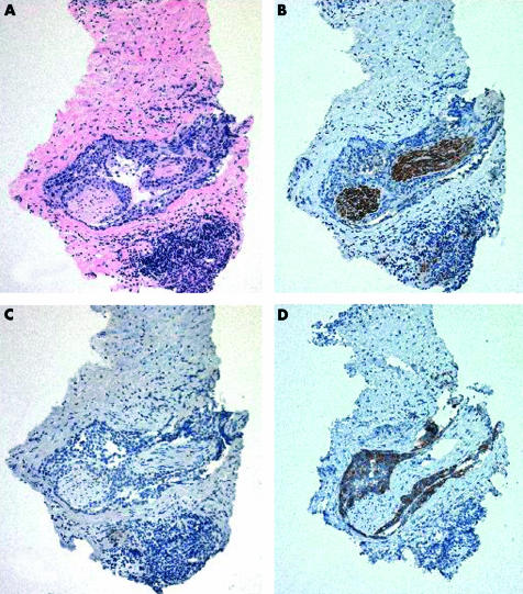
The diagnosis of prostatic adenocarcinoma remains dependent on the recognition of basic haematoxylin and eosin criteria. The discovery of alpha-methylacyl CoA racemase/P504S (AMACR/P504S) overexpression in prostate cancer represents a triumph of high throughput microarray technology, and is a powerful demonstration of how this methodology can be used to facilitate the rapid development of diagnostically relevant antibodies. Immunohistochemistry with anti-AMACR/P504S is useful for detecting prostate cancer in the full range of prostate specimens encountered in surgical pathology, be they needle biopsies, transurethral resection of prostate chips, or prostatectomies. In particular, studies to date with AMACR/P504S clearly demonstrate the ability of this marker to support a diagnosis of malignancy in prostate needle biopsies. This is particularly true when it is combined with negative staining for a basal cell marker, such as 34betaE12 or p63. Although it has limitations with respect to sensitivity and specificity, AMACR/P504S will no doubt become a standard adjunctive stain used by pathologists seeking to reach a definitive diagnosis in prostate biopsies considered to be atypical, but not diagnostic of malignancy on haematoxylin and eosin sections alone.
Figures


Similar articles
-
Expression and diagnostic utility of alpha-methylacyl-CoA-racemase (P504S) in foamy gland and pseudohyperplastic prostate cancer.Am J Surg Pathol. 2003 Jun;27(6):772-8. doi: 10.1097/00000478-200306000-00007. Am J Surg Pathol. 2003. PMID: 12766580
-
Detection of prostate cancer by alpha-methylacyl CoA racemase (P504S) in needle biopsy specimens previously reported as negative for malignancy.Histopathology. 2006 May;48(6):668-73. doi: 10.1111/j.1365-2559.2006.02409.x. Histopathology. 2006. PMID: 16681682
-
Diagnostic utility of a p63/alpha-methyl-CoA-racemase (p504s) cocktail in atypical foci in the prostate.Mod Pathol. 2004 Oct;17(10):1180-90. doi: 10.1038/modpathol.3800197. Mod Pathol. 2004. PMID: 15205683
-
Diagnostic utility of alpha-methylacyl CoA racemase (P504S) on prostate needle biopsy.Adv Anat Pathol. 2004 Nov;11(6):316-21. doi: 10.1097/01.pap.0000146924.14246.be. Adv Anat Pathol. 2004. PMID: 15505533 Review.
-
Discovery and clinical application of a novel prostate cancer marker: alpha-methylacyl CoA racemase (P504S).Am J Clin Pathol. 2004 Aug;122(2):275-89. doi: 10.1309/EJUY-UQPE-X1MG-68MK. Am J Clin Pathol. 2004. PMID: 15323145 Review.
Cited by
-
Alpha-methylacyl-CoA racemase (AMACR/P504S) protein expression in urothelial carcinoma of the upper urinary tract correlates with tumour progression.Virchows Arch. 2006 Mar;448(3):325-30. doi: 10.1007/s00428-005-0129-6. Epub 2005 Nov 29. Virchows Arch. 2006. PMID: 16315020
-
Molecular profiling of radical prostatectomy tissue from patients with no sign of progression identifies ERG as the strongest independent predictor of recurrence.Oncotarget. 2019 Nov 5;10(60):6466-6483. doi: 10.18632/oncotarget.27294. eCollection 2019 Nov 5. Oncotarget. 2019. PMID: 31741711 Free PMC article.
-
α-Methylacyl-CoA racemase spliced variants and their expression in normal and malignant prostate tissues.Urology. 2011 Jan;77(1):249.e1-7. doi: 10.1016/j.urology.2010.08.005. Urology. 2011. PMID: 21195844 Free PMC article.
-
Reactivation of androgen receptor-regulated lipid biosynthesis drives the progression of castration-resistant prostate cancer.Oncogene. 2018 Feb 8;37(6):710-721. doi: 10.1038/onc.2017.385. Epub 2017 Oct 23. Oncogene. 2018. PMID: 29059155 Free PMC article.
-
AMACR overexpression acts as a negative prognostic factor in oral squamous cell carcinoma.Int J Med Sci. 2018 Apr 3;15(6):638-644. doi: 10.7150/ijms.23291. eCollection 2018. Int J Med Sci. 2018. PMID: 29725255 Free PMC article.
References
-
- Thorson P, Humphrey PA. Minimal adenocarcinoma in prostate needle biopsy tissue. Am J Clin Pathol 2000;114:896–909. - PubMed
-
- Abrahams N, Ormsby A, Brainard J. Cytokeratin 5/6 is a basal cell-specific marker in benign prostatic glandular proliferations and in prostate tissue with androgen deprivation effect. Mod Pathol 2002;15:150A.
-
- Weinstein MH, Signoretti S, Loda M. Diagnostic utility of immunohistochemical staining for p63, a sensitive marker of prostatic basal cells. Mod Pathol 2002;15:1302–8. - PubMed
-
- Varma M, Linden MD, Amin MB. Effect of formalin fixation and epitope retrieval techniques on antibody 34betaE12 immunostaining of prostatic tissues. Mod Pathol 1999;12:472–8. - PubMed
-
- Oliai BR, Kahane H, Epstein JI. High molecular weight cytokeratin (HMWCK) reactivity in prostate carcinoma. Mod Pathol 2002;15:176A. - PubMed
Publication types
MeSH terms
Substances
LinkOut - more resources
Full Text Sources
Other Literature Sources
Medical