The cerebral oscillatory network of voluntary tremor
- PMID: 14645449
- PMCID: PMC1664802
- DOI: 10.1113/jphysiol.2003.051235
The cerebral oscillatory network of voluntary tremor
Erratum in
- J Physiol. 2004 May 1;556(Pt 3):1013
Abstract
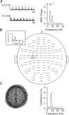
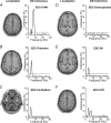
It has recently been shown that resting tremor in Parkinson's disease is associated with oscillatory neural coupling in an extensive cerebral network comprising a cerebello-diencephalic-cortical loop and cortical motor, somatosensory and posterior parietal areas contralateral to the tremor hand. The aim of the present study was to investigate whether this oscillatory brain network exclusively reflects a pathophysiological state in parkinsonian resting tremor or whether it constitutes a fundamental feature of physiological motor control. We investigated cerebro-muscular and cerebro-cerebral coupling in 11 healthy subjects imitating typical antagonistic parkinsonian tremor. We recorded brain activity with a 122-channel whole-head neuromagnetometer and surface EMGs of the forearm extensor. Analysis of cerebro-muscular and cerebro-cerebral coherence revealed oscillatory coupling in the same brain structures that comprise the oscillatory network of parkinsonian resting tremor. Interestingly, similar to parkinsonian resting tremor, cerebro-cerebral coherences often showed a significant peak at twice the simulated tremor frequency. The most striking differences between parkinsonian patients, as investigated in a previous study and healthy subjects imitating the antagonistic resting tremor were a reduction of the coupling between primary sensorimotor cortex and a diencephalic structure--most likely the thalamus--and an enhancement of the coupling between premotor and primary sensorimotor cortex. Our results indicate that the coupling of oscillatory activity within a cerebello-diencephalic-cortical loop constitutes a basic feature of physiological motor control. Thus, our data are consistent with the hypothesis that parkinsonian resting tremor involves oscillatory cerebro-cerebral coupling in a physiologically pre-existing network.
Figures


Similar articles
-
The cerebral oscillatory network of parkinsonian resting tremor.Brain. 2003 Jan;126(Pt 1):199-212. doi: 10.1093/brain/awg022. Brain. 2003. PMID: 12477707
-
Levodopa affects functional brain networks in Parkinsonian resting tremor.Mov Disord. 2009 Jan 15;24(1):91-8. doi: 10.1002/mds.22318. Mov Disord. 2009. PMID: 18823037
-
Pathological oscillatory coupling within the human motor system in different tremor syndromes as revealed by magnetoencephalography.Neurol Clin Neurophysiol. 2004 Nov 30;2004:26. Neurol Clin Neurophysiol. 2004. PMID: 16012624
-
Physiological and pathological oscillatory networks in the human motor system.J Physiol Paris. 2006 Jan;99(1):3-7. doi: 10.1016/j.jphysparis.2005.06.010. Epub 2005 Jul 27. J Physiol Paris. 2006. PMID: 16054347 Review.
-
The oscillating central network of Essential tremor.Clin Neurophysiol. 2012 Jan;123(1):61-4. doi: 10.1016/j.clinph.2011.09.024. Epub 2011 Nov 4. Clin Neurophysiol. 2012. PMID: 22055842 Review.
Cited by
-
Exertion dependent alternations in force fluctuation and limb acceleration during sustained fatiguing contraction.Eur J Appl Physiol. 2006 Jun;97(3):362-71. doi: 10.1007/s00421-006-0166-0. Epub 2006 May 10. Eur J Appl Physiol. 2006. PMID: 16770474
-
EEG Oscillations in Specific Frequency Bands Are Differently Coupled with Angular Joint Angle Kinematics during Rhythmic Passive Elbow Movement.Brain Sci. 2022 May 14;12(5):647. doi: 10.3390/brainsci12050647. Brain Sci. 2022. PMID: 35625033 Free PMC article.
-
Re-emergent Tremor in Parkinson's Disease: The Role of the Motor Cortex.Mov Disord. 2020 Jun;35(6):1002-1011. doi: 10.1002/mds.28022. Epub 2020 Mar 16. Mov Disord. 2020. PMID: 32175656 Free PMC article.
-
Towards a more robust non-invasive assessment of functional connectivity.Imaging Neurosci (Camb). 2024 Mar 28;2:imag-2-00119. doi: 10.1162/imag_a_00119. eCollection 2024. Imaging Neurosci (Camb). 2024. PMID: 40800450 Free PMC article.
-
The pace of prosodic phrasing couples the listener's cortex to the reader's voice.Hum Brain Mapp. 2013 Feb;34(2):314-26. doi: 10.1002/hbm.21442. Epub 2012 Jan 3. Hum Brain Mapp. 2013. PMID: 22392861 Free PMC article.
References
-
- Burkhard PR, Langston JW, Tetrud JW. Voluntarily simulated tremor in normal subjects. Neurophysiol Clin. 2002;32:119–126. - PubMed
-
- Deuschl G, Bain P, Brin M. Consensus statement of the Movement Disorder Society on Tremor. Ad Hoc Scientific Committee. Mov Disord. 1998;13:2–23. - PubMed
-
- Deuschl G, Raethjen J, Lindemann M, Krack P. The pathophysiology of tremor. Muscle Nerve. 2001;24:716–735. - PubMed
Publication types
MeSH terms
LinkOut - more resources
Full Text Sources
Other Literature Sources

