. 2003 Nov 26;23(34):10756-64.
doi: 10.1523/JNEUROSCI.23-34-10756.2003.

Mechanism of toxicity in rotenone models of Parkinson's disease

Affiliations

Mechanism of toxicity in rotenone models of Parkinson's disease

Todd B Sherer et al. J Neurosci. .

Abstract

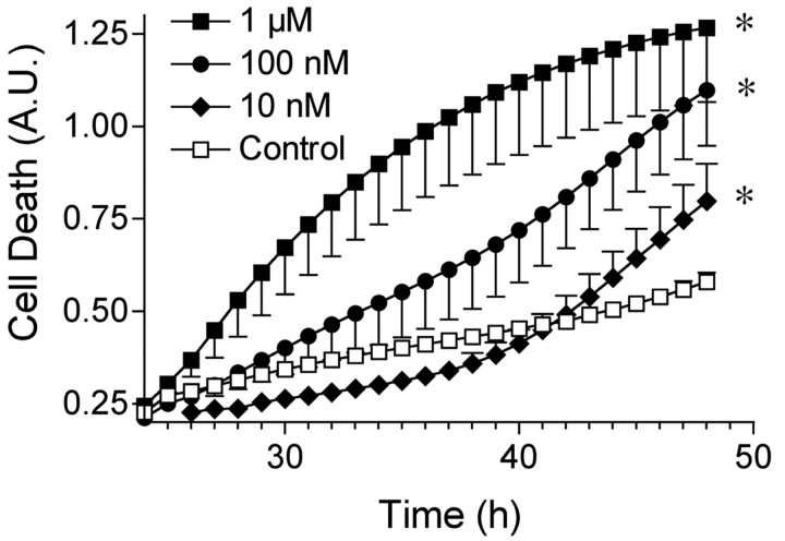
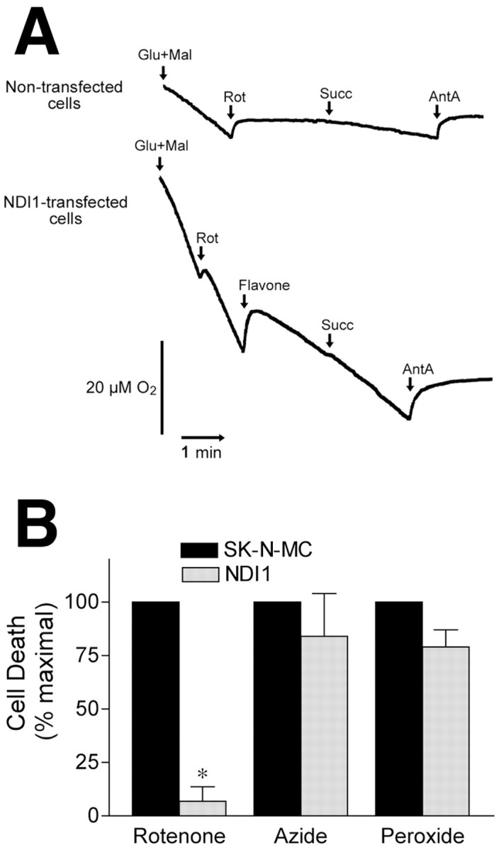
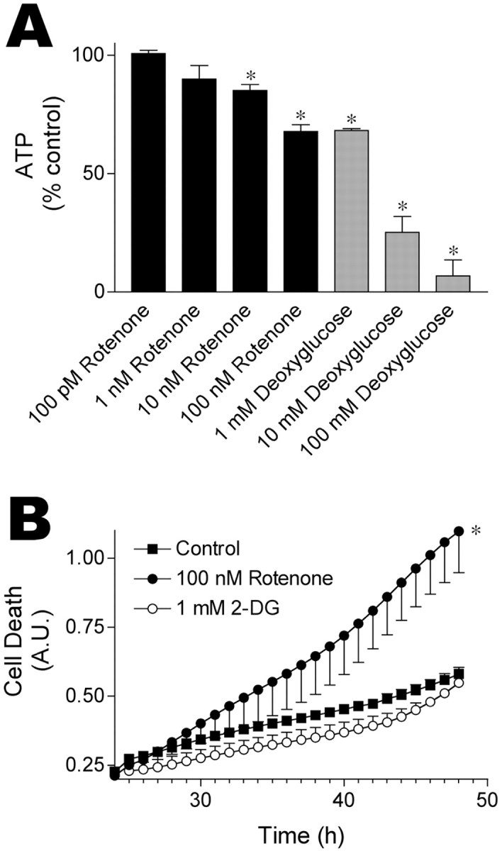
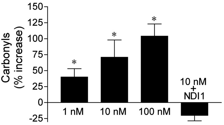
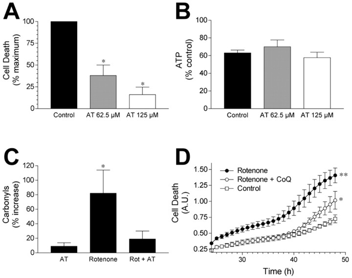
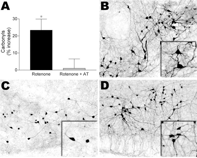
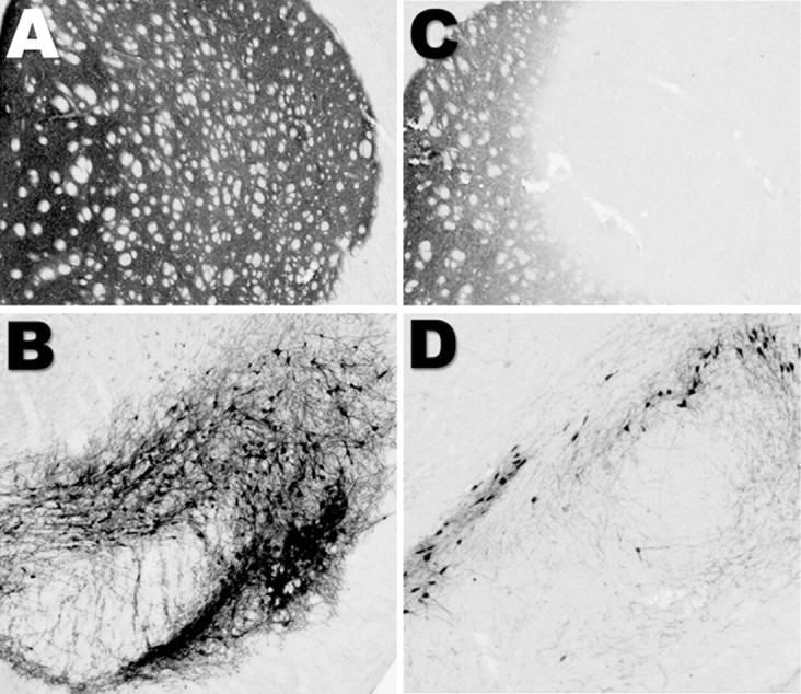
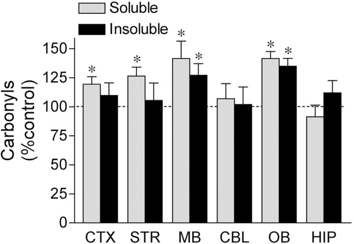
Exposure of rats to the pesticide and complex I inhibitor rotenone reproduces features of Parkinson's disease, including selective nigrostriatal dopaminergic degeneration and alpha-synuclein-positive cytoplasmic inclusions (Betarbet et al., 2000; Sherer et al., 2003). Here, we examined mechanisms of rotenone toxicity using three model systems. In SK-N-MC human neuroblastoma cells, rotenone (10 nm to 1 microm) caused dose-dependent ATP depletion, oxidative damage, and death. To determine the molecular site of action of rotenone, cells were transfected with the rotenone-insensitive single-subunit NADH dehydrogenase of Saccharomyces cerevisiae (NDI1), which incorporates into the mammalian ETC and acts as a "replacement" for endogenous complex I. In response to rotenone, NDI1-transfected cells did not show mitochondrial impairment, oxidative damage, or death, demonstrating that these effects of rotenone were caused by specific interactions at complex I. Although rotenone caused modest ATP depletion, equivalent ATP loss induced by 2-deoxyglucose was without toxicity, arguing that bioenergetic defects were not responsible for cell death. In contrast, reducing oxidative damage with antioxidants, or by NDI1 transfection, blocked cell death. To determine the relevance of rotenone-induced oxidative damage to dopaminergic neuronal death, we used a chronic midbrain slice culture model. In this system, rotenone caused oxidative damage and dopaminergic neuronal loss, effects blocked by alpha-tocopherol. Finally, brains from rotenone-treated animals demonstrated oxidative damage, most notably in midbrain and olfactory bulb, dopaminergic regions affected by Parkinson's disease. These results, using three models of increasing complexity, demonstrate the involvement of oxidative damage in rotenone toxicity and support the evaluation of antioxidant therapies for Parkinson's disease.


Figures

Figure 1.
Figure 1.
Dose-dependent cell death in response to rotenone exposure. Cells were exposed to rotenone for 48 hr, and cell death was monitored between 24 and 48 hr using Sytox green fluorescence. Rotenone treatment at 1 μm, 100 nm, and 10 nm caused toxicity. Results show means ± SEM of six independent experiments. *p < 0.05. A.U., Arbitrary units.
Figure 2.
Figure 2.
Rotenone toxicity requires electron transfer through complex I. A, Cells infected with rAAV-NDI1 are resistant to rotenone inhibition of oxygen utilization. Where indicated, 5 mm glutamate (Glu), 5 mm malate (Mal), 5 μm rotenone (Rot), 500 μm flavone, 5 mm succinate (Succ), and 5 μm antimycin (AntA) were added. Note that oxygen utilization in NDI1-transfected cells was not sensitive to rotenone inhibition but that antimycin A still inhibited oxygen consumption. B, NDI1-transduced cells were resistant to rotenone (100 nm) toxicity but remained sensitive to hydrogen peroxide (300 μm) and azide (1 mm). Control and NDI1-transduced cells were grown in 96 well plates, and cell death was analyzed 48 hr after exposure to compounds. Results represent means ± SEM for three independent experiments.
Figure 3.
Figure 3.
Rotenone toxicity does not result solely from ATP depletion. A, ATP depletion after exposure to rotenone or 2-DG. Note that rotenone (100 nm) and 2-DG (1 mm) caused similar reductions in cellular ATP levels. Cells were treated for 6-8 hr before ATP measurements; similar results were obtained at 20 hr. Data represent means ± SEM for three to five independent experiments. *p < 0.05 versus ATP levels in vehicle-treated wells. B, Although rotenone (100 nm) and 2-DG (1 mm) caused similar bioenergetic defects, only rotenone (100 nm) was toxic. Cells were exposed to rotenone or 2-DG for 48 hr, and cell death was determined using Sytox green fluorescence. Results show means ± SEM of three independent experiments, six replicates per experiment. *p < 0.05 versus death in vehicle-treated wells.
Figure 4.
Figure 4.
Rotenone-induced oxidative damage is dose-dependent and depends on electron transfer through complex I. Untransfected SK-N-MC and NDI1-transfected cells were exposed to rotenone for 24 hr, and then protein carbonyls were measured. Protein carbonyl levels are expressed as a percentage change from levels in solvent-treated cultures. NDI1-transfected cells received 10 nm rotenone for 24 hr. Data shown are means ± SEM and represent five to six independent experiments. *p < 0.05 versus solvent-treated controls.
Figure 5.
Figure 5.
Glutathione depletion potentiates rotenone toxicity. Cells were exposed to 10 μm BSO for 24 hr to deplete cellular glutathione before treatment with 10 nm rotenone. Cell death was then analyzed using Sytox green fluorescence, and death at 36 hr after rotenone treatment is shown. Results shown are means ± SEM for three independent experiments. *p < 0.05 versus solvent-treated controls; **p < 0.05 versus rotenone-treated wells.
Figure 6.
Figure 6.
Antioxidants protect cells from rotenone toxicity. A, α-Tocopherol pretreatment [62.5 μm (gray column) or 125 μm (white column)] attenuated rotenone toxicity. Death was expressed as a percentage of maximal cell death after rotenone treatment. Results represent means ± SEM for four to five independent experiments. *p < 0.05 versus rotenone-induced death (black column). B, α-Tocopherol pretreatment did not prevent ATP depletions after rotenone exposure. Cells were exposed to solvent or α-tocopherol (62.5 or 125 μm) before rotenone exposure, and ATP levels were measured. Column colors are as in A. Data shown are means ± SEM and represent three independent experiments. C, α-Tocopherol pretreatment prevented rotenone-induced oxidative damage. Cells were exposed to solvent or 125 μm α-tocopherol for 24 hr before rotenone (10 nm) treatment and before protein carbonyls were measured. Carbonyls are expressed as a percentage change from levels in solvent-treated cultures. Data shown are means ± SEM and represent five independent experiments. *p < 0.05 versus solvent-treated controls. D, Coenzyme Q10 (CoQ) pretreatment attenuated rotenone toxicity. Cells were pretreated for 24 hr with 12.5 μm coenzyme Q10 before exposure to 10 nm rotenone. Cell death was followed for 48 hr after rotenone treatment, and the last 24 hr period is shown. **p < 0.05 versus rotenone-treated cells in the presence of CoQ and versus solvent-treated cells; *p < 0.05 versus solvent-treated cells.
Figure 7.
Figure 7.
α-Tocopherol protected dopaminergic neurons in midbrain slice cultures from rotenone oxidative damage and toxicity. A, Rotenone caused oxidative damage to midbrain slice cultures that was prevented by α-tocopherol treatment. Slice cultures were exposed to rotenone (50 nm) for 7 d in the presence of vehicle or α-tocopherol (100 μm). Data are expressed as a percentage increase in protein carbonyl levels in rotenone-treated cultures. *p < 0.05 versus solvent-treated cells. B-D, Rotenone toxicity in midbrain slice cultures was prevented by α-tocopherol. Midbrain slice cultures were exposed to vehicle (B) or 50 nm rotenone for 7 d in the presence of vehicle (C) or α-tocopherol (100 μm) (D). Rotenone exposure markedly reduced the number of TH+ processes in midbrain dopaminergic neurons (compare B and C). α-Tocopherol treatment reduced the effects of rotenone on these degenerative changes (D).
Figure 8.
Figure 8.
Rotenone-induced loss of TH immunoreactivity in the nigrostriatal pathway. TH immunocytochemistry in the striatum (A) and substantia nigra (B) of a vehicle-treated rat is shown. Rotenone treatment reduced TH immunoreactivity in both the striatum (C) and the substantia nigra (D). The rotenone-treated animal received 3.0 mg/kg/d rotenone, subcutaneously, for 5 d.
Figure 9.
Figure 9.
Chronic rotenone infusion induced selective oxidative damage, with the greatest damage in the midbrain and the olfactory bulb. Chronic rotenone infusion increased protein carbonyl levels in soluble protein isolated from the striatum (n = 18), midbrain (n = 18), cortex (n = 11), and olfactory bulb (n = 12) but not the cerebellum (n = 10) or hippocampus (n = 10). Rotenone exposure increased insoluble protein carbonyl levels only in midbrain (n = 18) and olfactory bulb (n = 12). Data are expressed as a percentage of protein carbonyl levels in vehicle-treated animals. Results represent means ± SEM. *p < 0.05 compared with control.

Similar articles

Cited by

References

    1. Alam A, Schmidt WJ ( 2002) Rotenone destroys dopaminergic neurons and induces parkinsonian symptoms in rats. Behav Brain Res 136. - PubMed
    1. Alam ZI, Daniel SE, Lees AJ, Marsden DC, Jenner P, Halliwell B ( 1997) A generalised increase in protein carbonyls in the brain in Parkinson's but not incidental Lewy body disease. J Neurochem 69: 1326-1329. - PubMed
    1. Bai Y, Hajek P, Chomyn A, Chan E, Seo BB, Matsuno-Yagi A, Yagi T, Attardi G ( 2001) Lack of complex I activity in human cells carrying a mutation in MtDNA-encoded ND4 subunit is corrected by the Saccharomyces cerevisiae NADH-quinone oxidoreductase (NDI1) gene. J Biol Chem 276: 38808-38813. - PubMed
    1. Barrientos A, Moraes CT ( 1999) Titrating the effects of mitochondrial complex I impairment in the cell physiology. J Biol Chem 274: 16188-16197. - PubMed
    1. Betarbet R, Sherer TB, MacKenzie G, Garcia-Osuna M, Panov AV, Greenamyre JT ( 2000) Chronic systemic pesticide exposure reproduces features of Parkinson's disease. Nat Neurosci 3: 1301-1306. - PubMed

Publication types

MeSH terms

LinkOut - more resources