. 2003 Nov 26;23(34):10765-75.
doi: 10.1523/JNEUROSCI.23-34-10765.2003.

Progressive degradation and subsequent refinement of acoustic representations in the adult auditory cortex

Affiliations

Progressive degradation and subsequent refinement of acoustic representations in the adult auditory cortex

Shaowen Bao et al. J Neurosci. .

Abstract

Correlated neuronal activity is believed to play an important role in refining and maintaining cortical circuitry during early development. Here we provide evidence that globally and locally correlated activity mediate different forms of adult plasticity. Pulses of broad-spectrum noise were used to activate time-locked responses across large areas of the rat auditory cortex, globally synchronizing cortical activity. Brief tone pips were used to activate relatively small groups of neurons, generating locally correlated activity. Pairing pulsed noises with nucleus basalis (NB) stimulation in awake rats for 4 weeks broadened spectral tuning, disrupted tonotopic maps, and reduced spontaneous discharge correlation in the primary auditory cortex (AI), as examined under anesthesia. Those effects caused AI neurons to appear qualitatively similar to neurons in nonprimary auditory fields of naive animals. Subsequent pairing of tone pips with NB stimulation for a period of 4 weeks completely reversed these effects induced by previous noise-NB pairing. These findings further demonstrate that the adult auditory cortex retains a substantial capacity for receptive field plasticity and tonotopic map reorganization and that locally correlated activity plays an important role in plasticity in the adult, as in the developing cortex.


Figures

Figure 1.
Figure 1.
Pulsed noise evokes synchronized cortical activity. A, Cross-correlogram (-50 to 50 msec) constructed using spikes recorded during 3 sec periods of silence, pulsed noise, and pulsed tone. The frequency of the tone was at the CF of one of the neurons. The inset shows the frequency-intensity receptive fields of neurons sampled at the two sites. Noise but not tone synchronized activity of the two neurons. B, Total number of synchronized events as a function of distance. Spikes within 10 msec apart are consider as synchronized. Cross-correlograms from the pair of sites marked with the arrow are presented in A. C, Percentage of the synchronized events as functions of cortical distance. D, Z-score of neuronal discharge synchrony (see Materials and Methods) as a function of distance. In B-D, pulsed noise increased neuronal synchrony across all distances (i.e., open circles tend to be above closed circles). Pulsed tones increased synchrony when distances were short and decreased synchrony when distances were long (i.e., red circles tend to be above closed circles with distances <1 mm, and below closed circles with distances >1 mm). E, Shift-predictor-corrected number of synchronous events (i.e., the total number of synchronous events minus the number of synchronous events after one of the two spike trains was temporally shifted relative to the other), which is the number of synchronous events that were not accounted for by stimulus-locked responses. Shift-predictor-corrected synchrony was not enhanced by tone or noise stimulation, suggesting that the tone- or noise-evoked synchrony was caused by stimulus-locked responses. F, G, Number and percentage of synchronous events in a 600 msec window after NB stimulation. Similar to results in B and C, pulsed noise increased cortical synchrony across all distances, whereas tone pips increase synchrony only for sites <1 mm apart.
Figure 2.
Figure 2.
Degradation and refinement of primary auditory cortex by noise-NB pairing and subsequent tone-NB pairing. A-C, AI maps recorded from naive, noise-NB and noise-NB → tone-NB animals. Hatched regions had RF irregularity index scores >2. D-G, Representative receptive fields. The numbers in the parentheses are RF irregularity indices. Asterisks in B indicate sites tuned to 6.9 and 22.6 kHz. Scale bar, 1 mm.
Figure 6.
Figure 6.
Similarity between the properties of degraded AI and naive nonprimary auditory neurons. A, B, Representative cortical maps recorded from naive and noise-NB animals, showing both AI and nonprimary auditory fields. Dark lines outline the primary auditory cortex in the maps (o and x indicate sites nonresponsive to pure tones). C-H, Receptive fields recorded from sites 1-6 marked in A and B.
Figure 3.
Figure 3.
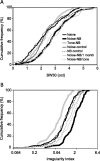
Quantitative degradation and refinement of the receptive fields of AI neurons. A, Noise-NB pairing broadened AI spectral tuning as indicated by increased response bandwidth at 30 dB above threshold. Subsequent pairing of NB stimulation with random tones (frequency of which was randomly chosen for each trial) sharpened the degraded spectral tuning. B, Noise-NB pairing significantly degraded AI receptive fields as measured with the RF irregularity index. Subsequent tone-NB pairing significantly improved RF. Noise experience (noise control) or NB stimulation (NB control) alone did not alter BW30 or RF irregularity index. The effects of noise-NB pairing were presented in animals examined 1 month after the last pairing procedure (noise-NB/1 month). One month of daily tone stimulation did not reverse noise-NB pairing effects.
Figure 4.
Figure 4.
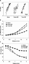
Quantitative degradation and refinement of the AI tonotopic maps. A, Rostral-caudal distribution of CFs along the AI tonotopic axis. B, Difference between CFs recorded from two sites as a function of the distance between the sites. C, Overlap of RFs of two sites as a function of the distance between the two sites. Note that noise-NB pairing disrupted AI tonotopicity, resulting in more scatted CF distribution, reduced CF difference at long distances, and increased RF overlap. Data from four noise-NB paired rats are shown in the graph for clarity. All six rats showed quantitatively similar effects. These degradation effects were reversed by subsequent tone-NB pairing.
Figure 5.
Figure 5.
Percentage of synchronized spontaneous discharge as a function of distance. Noise-NB pairing reduced neuronal synchrony. Subsequent tone-NB pairing reversed the effect.
Figure 7.
Figure 7.
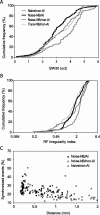
Similarity between properties of neurons in degraded AI and naive non-AI. A, Response bandwidth at 30 dB above threshold. B, A cumulative histogram of RF irregularity index. C, Percentage of synchronized spike events. Note that no significant differences were observed for these properties between the AI neurons of noise-NB paired animals and the nonprimary auditory neurons of naive animals.
Figure 8.
Figure 8.
Neuronal synchrony as a function of RF irregularity index score. Neuronal synchrony decreased with increasing RF irregularity.

References

    1. Ahissar E, Vaadia E, Ahissar M, Bergman H, Arieli A, Abeles M ( 1992) Dependence of cortical plasticity on correlated activity of single neurons and on behavioral context. Science 257: 1412-1415. - PubMed
    1. Arnold HM, Burk JA, Hodgson EM, Sarter M, Bruno JP ( 2002) Differential cortical acetylcholine release in rats performing a sustained attention task versus behavioral control tasks that do not explicitly tax attention. Neuroscience 114: 451-460. - PubMed
    1. Bakin JS, Weinberger NM ( 1996) Induction of a physiological memory in the cerebral cortex by stimulation of the nucleus basalis. Proc Natl Acad Sci USA 93: 11219-11224. - PMC - PubMed
    1. Bakin JS, South DA, Weinberger NM ( 1996) Induction of receptive field plasticity in the auditory cortex of the guinea pig during instrumental avoidance conditioning. Behav Neurosci 110: 905-913. - PubMed
    1. Bao S, Chan VT, Merzenich MM ( 2001) Cortical remodeling induced by activity of ventral tegmental dopamine neurons. Nature 412: 79-83. - PubMed

Publication types

LinkOut - more resources