. 2003 Nov 26;23(34):10800-8.
doi: 10.1523/JNEUROSCI.23-34-10800.2003.

Brain-derived neurotrophic factor-induced gene expression reveals novel actions of VGF in hippocampal synaptic plasticity

Affiliations

Brain-derived neurotrophic factor-induced gene expression reveals novel actions of VGF in hippocampal synaptic plasticity

Janet Alder et al. J Neurosci. .

Abstract

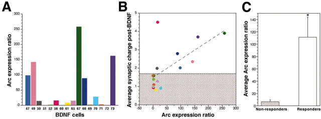
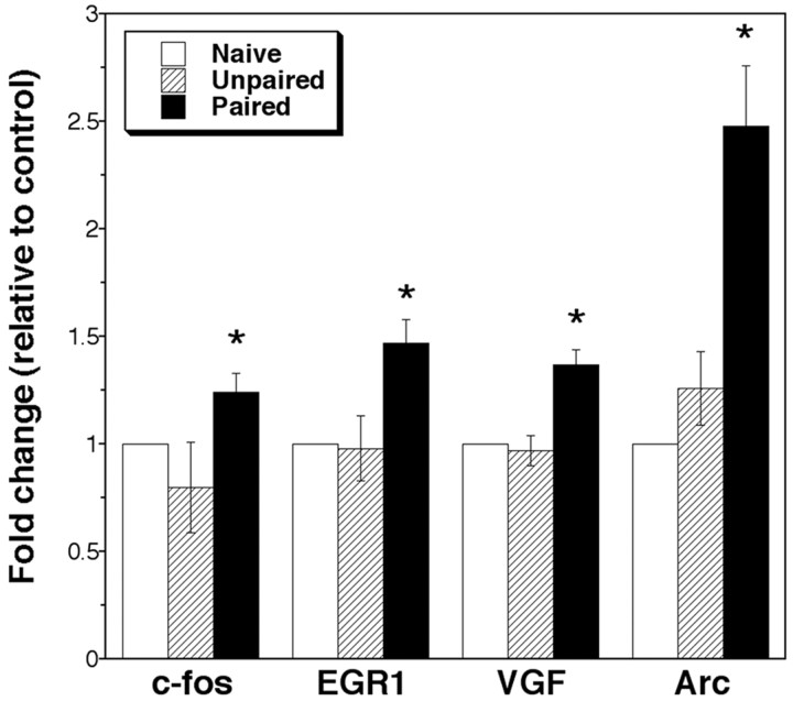
Synaptic strengthening induced by brain-derived neurotrophic factor (BDNF) is associated with learning and is coupled to transcriptional activation. However, identification of the spectrum of genes associated with BDNF-induced synaptic plasticity and the correlation of expression with learning paradigms in vivo has not yet been studied. Transcriptional analysis of BDNF-induced synaptic strengthening in cultured hippocampal neurons revealed increased expression of the immediate early genes (IEGs), c-fos, early growth response gene 1 (EGR1), activity-regulated cytoskeletal-associated protein (Arc) at 20 min, and the secreted peptide VGF (non-acronymic) protein precursor at 3 hr. The induced genes served as prototypes to decipher mechanisms of both BDNF-induced transcription and plasticity. BDNF-mediated gene expression was tyrosine kinase B and mitogen-activated protein kinase-dependent, as demonstrated by pharmacological studies. Single-cell transcriptional analysis of Arc after whole-cell patch-clamp recordings indicated that increased gene expression correlated with enhancement of synaptic transmission by BDNF. Increased expression in vitro predicted elevations in vivo: VGF and the IEGs increased after trace eyeblink conditioning, a hippocampal-dependent learning paradigm. VGF protein was also upregulated by BDNF treatment and was expressed in a punctate manner in dissociated hippocampal neurons. Collectively, these findings suggested that the VGF neuropeptides may regulate synaptic function. We found a novel function for VGF by applying VGF peptides to neurons. C-terminal VGF peptides acutely increased synaptic charge in a dose-dependent manner, whereas N-terminal peptide had no effect. These observations indicate that gene profiling in vitro can reveal new mechanisms of synaptic strengthening associated with learning and memory.


Figures

Figure 1.
Figure 1.
Hippocampal pyramidal-like neurons exhibit differential acute responses to BDNF. A, Example whole-cell voltage-clamp recordings (Vhold = -60 mV) from three individual hippocampal neurons during baseline recordings (NRS perfusion, -2 to 0 min) and 3-5 min after switching perfusion to either NRS (control) or BDNF (50 ng/ml). Note that cell 68 (blue) responds to BDNF, whereas cell 60 (red) does not. B, Synaptic charge in 1 min bins over a 20 min recording period for control neurons (average ± SE; n = 7) (thick black line), BDNF cell 68 (thick blue line), and BDNF cell 60 (thick red line) as well as all other individual neurons (all other colors; n = 16) exposed to BDNF. The differential response to BDNF is demonstrated. Recordings were obtained from multiple platings.
Figure 2.
Figure 2.
Arc expression and synaptic response to BDNF correlate at the single-cell level. A, Arc expression in individual BDNF-treated cells shown in Figure 1 after electrophysiological recordings and aRNA amplification. Arc transcription was measured by real time RT-PCR and then normalized to internal GAPDH levels. The data are then expressed as a ratio of the average Arc expression in the seven control cells (expression ratio). Colors for individual cells correspond to those in Figure 1. A large variability of Arc gene levels is apparent among the individual BDNF-treated cells. B, Correlation of Arc expression ratios to the synaptic BDNF responses of individual cells. Average synaptic charge 3-5 min after BDNF exposure expressed as a fold increase over baseline (-2 to 0 min) (Fig. 1) was plotted against Arc expression ratios shown in A. Each circle represents one BDNF-treated cell of corresponding color. Dashed line depicts a correlation coefficient indicating significance (r = 0.66; p < 0.005). The data are categorized into two groups of BDNF-treated cells: a cluster of non-responders (shaded) and a spread of responders (clear). C, Average Arc expression ratios of the two populations of cells defined in B. Responders (white bar) show a significantly higher fold increase in Arc expression (average ± SE; n = 7) compared with non-responders (shaded bar) (average ± SE; n = 9) (*p < 0.05; t test).
Figure 3.
Figure 3.
VGF as well as IEGs are upregulated after training with trace eyeblink conditioning. RNA was obtained from the hippocampi of naive rats, unpaired controls, and trace-conditioned rats after 800 trials (200 trials per day for 4 d) of paired stimuli and subjected to real time RT-PCR for a number of genes identified by microarray. Bars represent average gene expression ± SE for naive rats (n = 5; white bar), unpaired controls (n = 3; stippled bar), and trace conditioning (n = 8; black bar). All data are normalized to internal GAPDH levels and then expressed as a fold change over the amount of mRNA in naive (control) rats. A number of IEGs including c-fos, EGR1, and Arc were upregulated after trace conditioning. In addition the secreted polypeptide VGF was increased after paired trace conditioning. *Indicates paired samples are significantly different from unpaired and naive control samples (p < 0.05; ANOVA).
Figure 4.
Figure 4.
BDNF regulates the translation of the secreted polypeptide VGF. A, Western blot analysis of VGF expression after BDNF exposure (50 ng/ml). VGF protein appears as a doublet at 90 and 85 kDa. A minor high molecular weight species is detectable. B, Quantitation of VGF protein levels indicates that VGF is upregulated by 3 hr and is maximal at 12 hr. Bars represent average VGF expression (± SE; n = 2). Actin is used as an internal control for protein levels, and data are expressed as fold of VGF expression in untreated sister cultures. C, Immunocytochemical localization of VGF in dissociated hippocampal neurons 12 d in vitro. Both the soma and the neurites of the pyramidal-like neurons are positive for VGF expression. Scale bar, 50 μm.
Figure 5.
Figure 5.
Exogenous VGF peptides acutely enhance synaptic activity. A, Representative traces of whole-cell patch-clamp recordings (Vhold = -60 mV) on rat hippocampal cells during baseline recordings and 3-5 min after application of either the C-terminal VGF peptide (TLQP-62) or N-terminal VGF peptide (LEGS-28). B, TLQP-62 peptides (•) elicited an approximate 1.6-fold increase in synaptic charge within 2 min of exposure (indicated by horizontal bar) which remained elevated over the course of the recording (25 min). LEGS-28 peptides (○), however, did not affect synaptic charge for the duration of treatment. C, Dose-response effect of C-terminal VGF peptides (TLQP-62 and AQEE-30) compared with the response for the N-terminal peptide (LEGS-28). Maximal response is obtained at 0.1 μm for both TLQP-62 and AQEE-30. Average synaptic charge 3-5 min after VGF peptide perfusion relative to baseline (-2 to 0 min) is depicted (±SE). *Indicates significantly different from LEGS-28 (p < 0.05; ANOVA). Number of cells is shown in parentheses. Recordings were obtained from multiple platings.

References

    1. Adams JP, Sweatt JD ( 2002) Molecular psychology: roles for the ERK MAP kinase cascade in memory. Annu Rev Pharmacol Toxicol 42: 135-163. - PubMed
    1. Alonso M, Vianna MR, Depino AM, Mello e Souza T, Pereira P, Szapiro G, Viola H, Pitossi F, Izquierdo I, Medina JH ( 2002) BDNF-triggered events in the rat hippocampus are required for both short- and long-term memory formation. Hippocampus 12: 551-560. - PubMed
    1. Benson DL, Salton SR ( 1996) Expression and polarization of VGF in developing hippocampal neurons. Brain Res Dev Brain Res 96: 219-228. - PubMed
    1. Berninger B, Schinder AF, Poo MM ( 1999) Synaptic reliability correlates with reduced susceptibility to synaptic potentiation by brain-derived neurotrophic factor. Learn Mem 6: 232-242. - PMC - PubMed
    1. Beylin AV, Gandhi CC, Wood GE, Talk AC, Matzel LD, Shors TJ ( 2001) The role of the hippocampus in trace conditioning: temporal discontinuity or task difficulty? Neurobiol Learn Mem 76: 447-461. - PubMed

Publication types

MeSH terms

LinkOut - more resources