. 2003 Nov 26;23(34):10832-40.
doi: 10.1523/JNEUROSCI.23-34-10832.2003.

CaV1.3 channels are essential for development and presynaptic activity of cochlear inner hair cells

Affiliations

CaV1.3 channels are essential for development and presynaptic activity of cochlear inner hair cells

Andreas Brandt et al. J Neurosci. .

Abstract

Cochlear inner hair cells (IHCs) release neurotransmitter onto afferent auditory nerve fibers in response to sound stimulation. During early development, afferent synaptic transmission is triggered by spontaneous Ca2+ spikes of IHCs, which are under efferent cholinergic control. Around the onset of hearing, large-conductance Ca2+-activated K+ channels are acquired, and Ca2+ spikes as well as the cholinergic innervation are lost. Here, we performed patch-clamp measurements in IHCs of mice lacking the CaV1.3 channel (CaV1.3-/-) to investigate the role of this prevailing voltage-gated Ca2+ channel in IHC development and synaptic function. The small Ca2+ current remaining in IHCs from 3-week-old CaV1.3-/- mice was mainly mediated by L-type Ca2+ channels, because it was sensitive to dihydropyridines but resistant to inhibitors of non-L-type Ca2+ channels such as omega-conotoxins GVIA and MVIIC and SNX-482. Depolarization induced only marginal exocytosis in CaV1.3-/- IHC, which was solely mediated by L-type Ca2+ channels, whereas robust exocytic responses were elicited by photolysis of caged Ca2+. Secretion triggered by short depolarizations was reduced proportionally to the Ca2+ current, suggesting that the coupling of the remaining channels to exocytosis was unchanged. CaV1.3-/- IHCs lacked the Ca2+ action potentials and displayed a complex developmental failure. Most strikingly, we observed a continued presence of efferent cholinergic synaptic transmission and a lack of functional large-conductance Ca2+-activated K+ channels up to 4 weeks after birth. We conclude that CaV1.3 channels are essential for normal hair cell development and synaptic transmission.


Figures

Figure 4.
Figure 4.
Lack of functional large-conductance Ca2+-activated K+ channels in CaV1.3-/- IHC. A compares representative current traces of wt (P25) and mutant (P30) IHCs evoked by 150-msec-long step depolarizations to the indicated potentials. In B example traces of wt (-23 mV), wt + 5 mm TEA (-23 mV), and CaV1.3-/- (-25 mV) IHCs are shown on expanded time scale after normalizing them to their steady-state values reached at the end of the depolarization (outside axis, steady-state values were not reached within the displayed time frame, but see Fig. 4A) as well as in absolute numbers (inside axis). Normalized and absolute currents of wt overlap. C shows averaged current traces (mean ± SEM) obtained from CaV1.3-/- IHCs after depolarization to -8 mV, which had either low (no added Ca2+; 0.1 mm free EGTA in the pipette; n = 3 IHCs) or high (1 mm added Ca2+; no EGTA in the pipette; n = 5 IHCs) basal [Ca2+]i. For the latter, the change of intracellular [Ca2+]i was qualitatively confirmed by fura-2 fluorimetry (data not shown). D displays average I-V relationships for wt (n = 5 IHCs; P24-P29), wt + 5 mm TEA (n = 2 IHCs; P24, P29), and CaV1.3-/- (high Ca2+; n = 5 IHCs; P25) IHCs. “Early” and “late” designate the averaging time windows (shown as gray bars in A) of 1.2 msec for 100 μsec (mainly representing IK,f in wt) and 140 msec for 9.5 msec (representing the sum of all outward currents), respectively. E plots representative current density traces obtained from wt and mutant IHCs during a hyperpolarizing voltage step from -65 to -125 mV. The mean series resistance of all cells included in Figure 4 was 5.6 ± 0.3 MΩ after Rs compensation and data were off-line-corrected for the remaining voltage error.
Figure 1.
Figure 1.
Strongly reduced Ca2+ current and exocytosis in CaV1.3-/- IHCs. A plots representative Ca2+ current traces for wt (P31) and CaV1.3-/- (P32) IHCs, which were elicited by step depolarization to the peak Ca2+ current potential (Solutions Ii and Ie were used for A-D). Data were leak-subtracted using a p/6 protocol. In B average current-voltage relationships (I-V) recorded from wt (n = 6 cells) and a CaV1.3-/- (n = 7 cells) IHCs are displayed. The dashed line represents a scaled version of the CaV1.3-/-I-V (scaling factor of 12.8 to match peak Ca2+ currents). C, D, Capacitance responses (ΔCm) to paired depolarizations to the peak Ca2+ current potential, spaced 100 msec apart and having variable duration of 10, 20, 50, and 100 msec were recorded for wt (n = 6 cells; P20-P32) and CaV1.3-/- (n = 9 cells; P14-P32) IHCs. C shows representative examples of wt and CaV1.3-/- ΔCm in response to a 2 * 20 msec paired pulse (low-pass filtered at 100 Hz). The bars indicate averages of baseline-subtracted CaV1.3-/-Cm over 400 msec before and 40-100 msec after the first and second depolarization. Intervals between the pairs were at least 30 sec to allow for complete recovery of the readily releasable pool (Moser and Beutner, 2000). D plots mean wt and mutant ΔCm responses to the first depolarization versus the respective duration of the stimuli. E shows the average ΔCm response of CaV1.3-/- IHCs (n = 9 recordings) to flash-photolysis of caged Ca2+. The mean CaV1.3-/- ΔCm is displayed along with its SE.
Figure 2.
Figure 2.
Developmental changes of Ca2+ current density: lack of Ca2+ spikes in CaV1.3-/- IHCs. A shows mean absolute (top panel) and normalized (bottom panel, normalized to P6 values) Ca2+ current densities for CaV1.3-/-, C57BL/6J, and NMRI IHCs (the NMRI data set was taken from Beutner and Moser, 2001) at P6 (nNMRI = 10; nC57BL/6J = 2; nCav1.3-/- = 5 IHCs), P10 (nNMRI = 9; nCav1.3-/- = 7 IHCs), and P14-P32 (nNMRI = 28; nC57BL/6J = 8; nCav1.3-/- = 8 IHCs). The top panel highlights the dramatic reduction in Ca2+ current density in CaV1.3-/- IHCs. The bottom panel emphasizes the developmental current density reduction in all strains investigated. B, Whereas current injection readily triggered APs in wt IHCs (left panel, example is P5), even strong current injections failed to elicit APs in CaV1.3-/- IHCs (right panel, example is P7). The inset compares the initial rise of membrane potential of representative wt and CaV1.3-/- IHCs after injection of 300 pA.
Figure 3.
Figure 3.
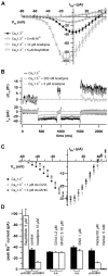
Identity and function of the Ca2+ channels in CaV1.3-/- IHCs. A plots average Ba2+ current IVs (mean ± SEM) of mutant IHCs under control conditions and during application of the DHP agonist BayK8644 (5 μm; n = 4 cells), the DHP antagonist isradipine (10 μm; n = 7 cells), or nickel (5 mm; n = 2 cells). Control I-V relationships before application of DHP agonists and antagonists were pooled. Individual I-V relationships were leak-corrected (see Materials and Methods). B compares leak-corrected Ca2+ currents and corresponding ΔCm recorded in the perforated-patch configuration in response to pairs of 500 msec depolarizations to -6 mV during subsequent treatment with 200 nm and 5 μm of isradipine. C plots average Ba2+ current I-V relationships of a representative perforated-patch experiment in which application of 1 μm of ω-conotoxin (ctx) GVIA was followed by wash-in of 3 μm of ω-conotoxin MVIIC without any obvious effects on current amplitude or voltage dependence. D summarizes the mean effects of the specified drugs on the CaV1.3-/- IHCs peak Ba2+ current. Each group of drugs was compared with their own control data acquired in perforated-patch experiments before the wash-in of the drug. The error probabilities for assuming statistically different mean amplitudes are provided underneath the plot.
Figure 5.
Figure 5.
Continued presence of postsynaptic responses in CaV1.3-/- IHCs. A compares representative spontaneous postsynaptic currents of wt and CaV1.3-/- IHCs before and after the onset of hearing in wt (potentials are indicated next to the traces). I-V relationships of the slow component of the postsynaptic currents are plotted in B. C displays biphasic postsynaptic currents of a P30 CaV1.3-/- IHC (at -75 mV) in more detail. D shows a stretch of a current-clamp Vm recording from the same cell as in C, illustrating the depolarizing onset and hyperpolarizing main component of these postsynaptic potentials. Extracellular application of 300 nm strychnine (P35; E) and dequalinium (1 μm; F; same cell as in E) abolished the postsynaptic currents of CaV1.3-/- IHCs. The inhibitory effects of both drugs were reversible.

References

    1. Art JJ, Fettiplace R ( 1987) Variation of membrane properties in hair cells isolated from the turtle cochlea. J Physiol (Lond) 385: 207-242. - PMC - PubMed
    1. Bech-Hansen NT, Naylor MJ, Maybaum TA, Pearce WG, Koop B, Fishman GA, Mets M, Musarella MA, Boycott KM ( 1998) Loss-of-function mutations in a calcium-channel alpha1-subunit gene in Xp11.23 cause incomplete X-linked congenital stationary night blindness. Nat Genet 19: 264-267. - PubMed
    1. Beutner D, Moser T ( 2001) The presynaptic function of mouse cochlear inner hair cells during development of hearing. J Neurosci 21: 4593-4599. - PMC - PubMed
    1. Beutner D, Voets T, Neher E, Moser T ( 2001) Calcium dependence of exocytosis and endocytosis at the cochlear inner hair cell afferent synapse. Neuron 29: 681-690. - PubMed
    1. Brown AM, Kunze DL, Yatani A ( 1984) The agonist effect of dihydropyridines on Ca channels. Nature 311: 570-572. - PubMed

Publication types

MeSH terms

Substances