. 2003 Nov 26;23(34):10913-22.
doi: 10.1523/JNEUROSCI.23-34-10913.2003.

Long-lasting functional disabilities in middle-aged rats with small cerebral infarcts

Affiliations

Long-lasting functional disabilities in middle-aged rats with small cerebral infarcts

Mark D Lindner et al. J Neurosci. .

Abstract

Recommendations from experts and recently established guidelines on how to improve the face and predictive validity of animal models of stroke have stressed the importance of using older animals and long-term behavioral-functional endpoints rather than relying almost exclusively on acute measures of infarct volume in young animals. The objective of the present study was to determine whether we could produce occlusions in older rats with an acceptable mortality rate and then detect reliable, long-lasting functional deficits. A reversible intraluminar suture middle cerebral artery occlusion (MCAO) procedure was used to produce small infarcts in middle-aged rats. This resulted in an acceptable mortality rate, and robust disabilities were detected in functional assays, although the degree of total tissue loss measured 90 d after MCAO was quite modest. Infarcted animals were functionally impaired relative to sham control animals even 90 d after the occlusions, and when animals were subgrouped based on amount of tissue loss, MCAO animals with only 4% tissue loss exhibited enduring neurological-behavioral impairments relative to sham-operated controls, and the functional impairments in the group with the largest infarcts (20% tissue loss) were more severe than the functional impairments in the rats with 4% tissue loss. These results suggest that this model, using reversible MCAO to produce small infarcts and long-lasting functional-behavioral deficits in older rats, may represent an advance in the relatively higher-throughput modeling of stroke and its recovery in rodents and may be useful in the development and characterization of future stroke therapies.


Figures
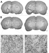
Figure 1.
Figure 1.
Representative photomicrographs of infarcts of the 20% tissue loss (A) and 4% tissue loss (B) groups. The infarcted area is indicated by the thick black lines in A and the arrows in B. The thinner black lines delineate the division between the hemispheres. Each section is from a different animal and is representative of the infarct at that coronal plane. Numbers indicate approximate coordinates in millimeters anterior to the interaural line. Scale bar, 1 mm. CTX, Cortex; STR, striatum. C, Higher-magnification view of the region of striatal infarct shown in B reveals clear gliosis and no definable neurons. D, A comparable region in the contralateral striatum showing a normal mixture of glia and neurons. Scale bar, 50 μm.
Figure 2.
Figure 2.
Volumetric measures of cortex, striatum, and the entire hemisphere for rats in the control group (CTL; n = 6), rats with 4% tissue loss of the entire hemisphere (n = 16), and rats with 20% tissue loss of the entire hemisphere (n = 6). A, Volumes of neocortex in hemisphere ipsilateral and contralateral to occlusion. B, Volumes of striatum in hemispheres ipsilateral and contralateral to occlusion. C, Volumes of entire hemisphere ipsilateral and contralateral to occlusion. Volumes of occluded structures that are significantly smaller than the contralateral side are indicated by asterisks. Data are presented in text and figures as means ± SEM.
Figure 3.
Figure 3.
Tests discontinued 30 d after surgery because of insufficient sensitivity to MCA occlusions. A, Bracing, which is the number of postural adjustments with affected forelimb while being pushed sideways for 90 cm. B, Akinesia, which is the number of steps initiated spontaneously with the affected forelimb during 30 sec trials. C, Rotorod, which is latency to fall from accelerating rotorod (5-40 rpm during 60 sec trial). Control rats (n = 9), rats with 4% tissue loss (n = 18), and rats with 20% tissue loss (n = 6) are shown.
Figure 4.
Figure 4.
Performance on tests of forelimb function that were sensitive to MCAO. A, Forelimb adduction. B, Tactile adhesive test of forelimb somatosensory function. C, Placing test of forelimb somatomotor function. D, Staircase test of forelimb and paw fine motor control. E, Fixed-ratio bar pressing test of forelimb motor speed and endurance. Control rats (n = 9), rats with 4% tissue loss (n = 18), and rats with 20% tissue loss (n = 6) are shown.
Figure 5.
Figure 5.
Composite behavioral score combining the five behavioral measures in Figure 4.A, All MCAO rats combined. B, MCAO rats divided into subgroups with 4% tissue loss and 20% tissue loss. Deficits are evident on day 1 even in the sham occlusion group. Both occluded groups are significantly impaired throughout the 90 d testing period. *Significantly different from control. **Significantly different from 4% tissue loss group.
Figure 6.
Figure 6.
Volumetric measures of cortex, striatum, and entire hemisphere for rats grouped by occlusion duration: Control group (CTL; n = 6), rats with 1 hr occlusions (n = 12), and rats with 2 hr occlusions (n = 10) are shown. A, Volumes of neocortex in hemisphere ipsilateral and contralateral to occlusion. B, Volumes of striatum in hemispheres ipsilateral and contralateral to occlusion. C, Volumes of entire hemisphere ipsilateral and contralateral to occlusion. Volumes of occluded structures that are significantly smaller than the contralateral side are indicated with asterisks.
Figure 7.
Figure 7.
Composite behavioral scores with groups on the basis of occlusion duration combining the five behavioral measures that were sensitive to the infarcts. Both occluded groups were significantly impaired throughout the 90 d testing period. *Significantly different from control. **Significantly different from 1 hr occlusion group.

Similar articles

Cited by

References

    1. Abrous DN, Dunnett SB ( 1994) Paw reaching in rats: the staircase test. In: Neuroscience protocols, module 3 (Wouterlood FG, ed), pp 19-29. Amsterdam: Elsevier.
    1. American Heart Association ( 2001) Heart and stroke statistical update, 2000. Dallas: American Heart Association.
    1. Andersen MB, Zimmer J, Sams-Dodd F ( 1999) Specific behavioral effects related to age and cerebral ischemia in rats. Pharmacol Biochem Behav 62: 673-682. - PubMed
    1. Belayev L, Alonso OF, Busto R, Zhao WZ, Ginsberg MD ( 1996) Middle cerebral artery occlusion in the rat by intraluminal suture: neurological and pathological evaluation of an improved model. Stroke 27: 1616-1622. - PubMed
    1. Bland ST, Schallert T, Strong R, Aronowski J, Grotta JC, Feeney DM ( 2000) Early exclusive use of the affected forelimb after moderate transient focal ischemia in rats: functional and anatomic outcome. Stroke 31: 1144-1152. - PubMed

LinkOut - more resources