. 2003 Nov 26;23(34):10934-43.
doi: 10.1523/JNEUROSCI.23-34-10934.2003.

Neurosteroids shift partial agonist activation of GABA(A) receptor channels from low- to high-efficacy gating patterns

Affiliations

Neurosteroids shift partial agonist activation of GABA(A) receptor channels from low- to high-efficacy gating patterns

Matt T Bianchi et al. J Neurosci. .

Abstract

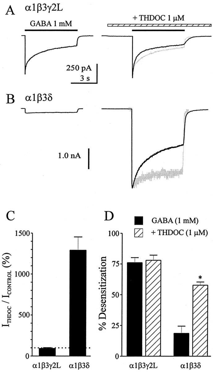
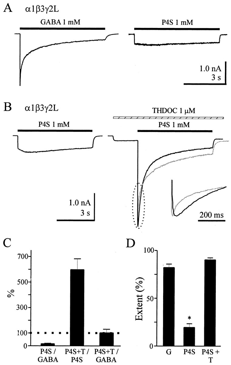
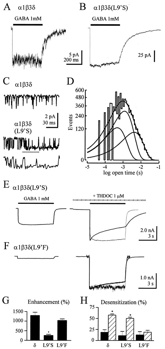
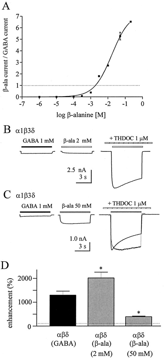
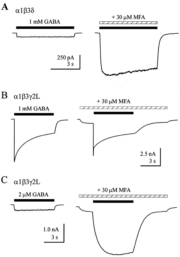
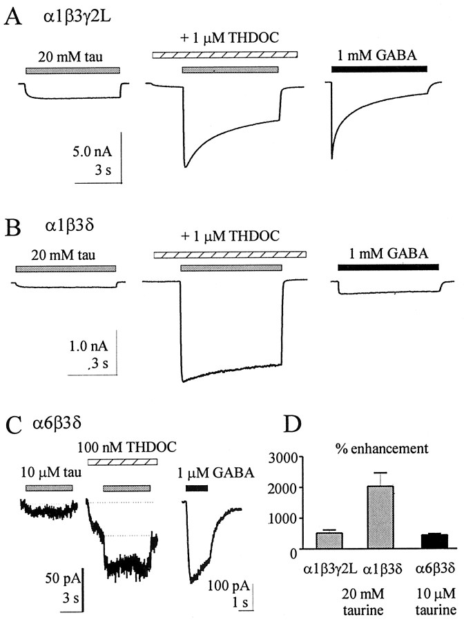
Although GABA activates synaptic (alphabetagamma) GABA(A) receptors with high efficacy, partial agonist activation of alphabetagamma isoforms and GABA activation of the primary extrasynaptic (alphabetadelta) GABA(A) receptors are limited to low-efficacy activity, characterized by minimal desensitization and brief openings. The unusual sensitivity of alphabetadelta receptor channels to neurosteroid modulation prompted investigation of whether this high sensitivity was dependent on the delta subunit or the low-efficacy channel function that it confers. We show that the isoform specificity (alphabetadelta > alphabetagamma) of neurosteroid modulation could be reversed by conditions that reversed isoform-specific activity modes, including the use of beta-alanine to achieve increased efficacy with alphabetadelta receptors and taurine to render alphabetagamma receptors low efficacy. We suggest that neurosteroids preferentially enhance low-efficacy GABA(A) receptor activity independent of subunit composition. Allosteric conversion of partial to full agonism may be a general mechanism for reversibly scaling the efficacy of GABA(A) receptors to endogenous partial agonists.


Figures

Figure 1.
Figure 1.
THDOC differentially modulated α1β3γ2L and α1β3δ GABAA receptor currents. A, α1β3γ2L GABAA receptor currents evoked by GABA (1 mm; left trace) were minimally affected by pre-applied THDOC (1 μm; right trace). In this and subsequent figures, GABA applications are indicated by solid bars, and THDOC applications are indicated by hatched bars. B, α1β3δ GABAA receptor currents evoked by GABA (1 mm; left trace) were markedly enhanced by pre-applied THDOC (1 μm; left trace). The time calibration is the same as in A. In A and B, the GABA alone trace was scaled to peak and overlaid (gray trace) for comparison of the time course. C, THDOC enhancement of peak current amplitude for α1β3γ2L and α1β3δ GABAA receptors is summarized. The dotted line indicates 100%. D, The extent of desensitization measured with GABA alone (solid bars) and with THDOC (hatched bars) is compared. Data are mean ± SEM. *p < 0.05, compared with GABA alone condition.
Figure 2.
Figure 2.
α1β3γ2L GABAA receptor currents evoked by the low-efficacy agonist P4S were enhanced by THDOC. A, Current traces recorded from the same cell expressing α1β3γ2L GABAA receptors in response to 1 mm GABA (left) or 1 mm P4S, a low-efficacy GABAA receptor agonist (right). B, α1β3γ2L receptor currents evoked by P4S (solid bar, left) were markedly enhanced by pre-applied THDOC (hatched bar, right). The response to GABA alone (1 mm) in the same cell is scaled and overlaid (gray) with the P4S plus THDOC current for comparison. The first 200 msec (indicated by the dotted oval) was expanded in the inset. C, Comparison of currents evoked by GABA, P4S, and P4S plus THDOC. For each cell, the P4S amplitude was <20% of the GABA-evoked current (left bar), the P4S plus THDOC amplitude was ∼600% of the P4S amplitude (middle bar), and the P4S plus THDOC amplitude was not different from the GABA alone amplitude (right bar). The dotted line indicates 100%. D, The extent of desensitization is shown for currents evoked by GABA alone (G), P4S alone, and P4S plus THDOC (T). Asterisk indicates significant difference from GABA alone and GABA plus THDOC (p < 0.0001).
Figure 3.
Figure 3.
α1β3δ (L9'S) GABAA receptors exhibited increased gating efficacy. A, B, Concentration-jump experiments (400 msec) performed on outside-out patches demonstrated minimal desensitization in response to GABA (1 mm) for α1β3δ (A) and α1β3δ (L9'S) (B) isoforms. C, Representative single-channel records are presented from α1β3δ (top trace) and α1β3δ (L9'S) receptors. A portion of the middle trace, indicated by the open bar, is expanded in the bottom trace. D, Event histogram of α1β3δ (L9'S) receptor single-channel openings evoked by steady state application of GABA (1 mm). Data were pooled from two patches. The distribution was best described by the sum of three exponential functions. The individual fitted functions, as well as their sum, are shown as curves on the plot. See Results for time constants and relative areas. E, F, Currents evoked by GABA (1 mm; solid bar) applied to cells expressing α1β3δ (L9'S) (E) and α1β3δ (L9′F) (F) in the absence (left traces) and presence (right traces) of THDOC (1 μm; hatched bar). G, Extent of desensitization (percentage) for 6 sec applications of GABA in the absence (solid bars) or presence of pre-applied THDOC (1 μm; hatched bars).
Figure 4.
Figure 4.
Attenuated neurosteroid enhancement of α1β3δ receptor currents evoked by high, but not low, concentrations of β-alanine. A, Concentration-response relationship for α1β00 receptor currents evoked by β-alanine. Currents were normalized to the amplitude of a maximal GABA response evoked in each cell by 1 mm GABA (dotted line). Data were from three cells. B, Currents evoked by GABA (1 mm; left trace) and β-alanine (2 mm; middle trace) from the same cell, as well as the enhancement of β-alanine current by pre-applied THDOC (1 μm; right trace). C, Same protocol as in B, except that 50 mm β-alanine was used, a concentration that evoked currents approximately fivefold larger than those evoked by GABA. D, Summary of THDOC enhancement of peak current under various conditions. Asterisk indicates significant difference from THDOC enhancement of currents evoked by GABA (1 mm).
Figure 5.
Figure 5.
The nonsteroidal ibuprofen analog MFA differentially modulated α1β3d and α1β3γ2L GABAA receptor currents. A, α1β3δ receptor currents evoked by 1 mm GABA alone (left trace, solid bar) were markedly enhanced by co-applied 30 μm MFA (hatched bar, right trace). B, α1β3γ2L receptor currents evoked by 1 mm GABA alone (left) or with pre-applied 30 μm MFA (right). C, α1β3γ2L receptor currents evoked by 2 μm GABA were potentiated by 30 μm MFA. Similar results were obtained in at least three additional cells for each condition.
Figure 6.
Figure 6.
THDOC enhancement of GABAA receptor currents evoked by the endogenous partial agonist taurine. A, α1β3γ2L receptor current evoked by 20 mm taurine alone (solid bar, left trace) or with pre-applied 1 μm THDOC (hatched bar, middle trace). The maximal GABA-evoked current is shown in the same cell for comparison (1 mm; solid bar, right trace). Scale bars apply to all three traces, and the time bar applies to other panels (except C, right trace). B, α1β3δ receptor current evoked by 20 mm taurine alone (solid bar, left trace) or with pre-applied 1 μm THDOC (hatched bar, middle trace). The maximal current evoked by 1 mm GABA is shown from the same cell (solid bar, right trace). C, α6β3δ receptor current evoked by 10 μm taurine alone (solid bar, left trace) or with pre-applied 100 nm THDOC (hatched bar, middle trace). The response to 1 μm GABA from the same cell is shown (right trace; note the different horizontal and vertical scale bars). The dotted line (left trace) is a baseline reference for the small taurine-evoked current. D, Summary plot indicating peak current enhancement by THDOC for the various conditions (n = 4 cells for each).

Similar articles

Cited by

References

    1. Adkins CE, Pillai GV, Kerby J, Bonnert TP, Haldon C, McKernan RM, Gonzalez JE, Oades K, Whiting PJ, Simpson PB ( 2001) α4β3δ GABAA receptors characterized by fluorescence resonance energy transfer-derived measurements of membrane potential. J Biol Chem 276: 38934-38939. - PubMed
    1. Angelotti TP, Uhler MD, Macdonald RL ( 1993) Assembly of GABAA receptor subunits: analysis of transient single-cell expression utilizing a fluorescent substrate/marker gene technique. J Neurosci 13: 1418-1428. - PMC - PubMed
    1. Bai D, Zhu G, Pennefather P, Jackson MF, MacDonald JF, Orser BA ( 2001) Distinct functional and pharmacological properties of tonic and quantal inhibitory postsynaptic currents mediated by gamma-aminobutyric acid(A) receptors in hippocampal neurons. Mol Pharmacol 59: 814-824. - PubMed
    1. Bianchi MT, Macdonald RL ( 2001) Mutation of the 9′ leucine in the GABAA receptor γ2L subunit produces an apparent decrease in desensitization by stabilizing open states without altering desensitized states. Neuropharmacology 41: 737-744. - PubMed
    1. Bianchi MT, Haas KF, Macdonald RL ( 2002) alpha1 and alpha6 subunits specify distinct desensitization, deactivation and neurosteroid modulation of GABA(A) receptors containing the delta subunit. Neuropharmacology 43: 492-502. - PubMed

Publication types

MeSH terms

LinkOut - more resources