. 2003 Dec;23(24):9127-35.
doi: 10.1128/MCB.23.24.9127-9135.2003.

Calpain 3 is activated through autolysis within the active site and lyses sarcomeric and sarcolemmal components

Affiliations

Calpain 3 is activated through autolysis within the active site and lyses sarcomeric and sarcolemmal components

Mathieu Taveau et al. Mol Cell Biol. 2003 Dec.

Abstract

Calpain 3 (Capn3) is known as the skeletal muscle-specific member of the calpains, a family of intracellular nonlysosomal cysteine proteases. This enigmatic protease has many unique features among the calpain family and, importantly, mutations in Capn3 have been shown to be responsible for limb girdle muscular dystrophy type 2A. Here we demonstrate that the Capn3 activation mechanism is similar to the universal activation of caspases and corresponds to an autolysis within the active site of the protease. We undertook a search for substrates in immature muscle cells, as several lines of evidence suggest that Capn3 is mostly in an inactive state in muscle and needs a signal to be activated. In this model, Capn3 proteolytic activity leads to disruption of the actin cytoskeleton and disorganization of focal adhesions through cleavage of several endogenous proteins. In addition, we show that titin, a previously identified Capn3 partner, and filamin C are further substrates of Capn3. Finally, we report that Capn3 colocalizes in vivo with its substrates at various sites along cytoskeletal structures. We propose that Capn3-mediated cleavage produces an adaptive response of muscle cells to external and/or internal stimuli, establishing Capn3 as a muscle cytoskeleton regulator.


Figures
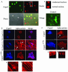
FIG. 1.
FIG. 1.
Capn3 ectopic expression leads to cell rounding and nuclear condensation associated with disruption of the actin stress fibers and disorganization of focal adhesions. (A) NIH 3T3 mouse fibroblasts were transiently transfected with pTOM-Capn3, encoding Capn3 tagged with two eGFP variants. Images were visualized by confocal microscopy. Transfected cells appear in green. Nuclei were stained with Topro 3 (red). Insert a shows examples of nuclear condensation in transfected cells and the shape of the nucleus in a nontransfected cell. White arrows in the phase image point to round cells. Control cells were transfected with pTOM. Bar = 10 μm. (B) Mouse myoblasts were transiently transfected with pSRD-Capn3 or pSRD-Capn3C129S as a control. Transfected cells were stained with anti-Capn3 RP1. F-actin was labeled with phalloidin. Images were visualized by confocal microscopy. Two examples of pSRD-Capn3 transfected cells stained for actin are shown. Insert a shows actin aggregates in the cytoplasm of transfected cells. Insert b shows actin fibers in a nontransfected cell. Bar = 10 μm. (C) Mouse myoblasts were transiently transfected with pSRD-Capn3 or pSRD-Capn3C129S as a control. Transfected cells were stained with anti-Capn3 RP1. Focal adhesions were stained with a monoclonal anti-alpha actinin antibody. Images were visualized by confocal microscopy. Insert a shows loss of the focal adhesion rod shape, and insert b shows normal rod-shaped focal adhesions. Bar = 10 μm.
FIG. 2.
FIG. 2.
Capn3 is activated through autolysis in the active site which involves intra- and intermolecular events. (A) Mouse myoblasts were transiently transfected with pSRD-Capn3ΔExon6 or pSRD-Capn3Y274A or cotransfected with plasmids coding for the 34-kDa autolysis fragment (pDEST40-Capn334-274) and the 55-kDa autolysis fragment (pDEST47-Capn3323-821). Images were visualized by confocal microscopy. Capn3 was detected with anti-Capn3 RP1. Capn334-274 was detected with a monoclonal anti-V5 tag and Capn3323-821 was detected with eGFP. Actin was labeled with phalloidin and focal adhesions were stained with a monoclonal anti-alpha actinin antibody or a monoclonal anti-vinculin antibody. Bar = 10 μm. (B) Schematic representation of Capn3 proteolytic domain with the C129S, Y274A, and Y322A mutations indicated in green, red, and blue, respectively. (C) 911 cells were transfected with pSRD-wt-Capn3, pSRD-Capn3C129S, pSRD-Capn3Y274A, and pSRD-Capn3Y322A alone or in combination. Twenty-four hours after transfection, cells were harvested and lysed for protein extraction. Western blot analysis of Capn3 was performed with anti-Capn3 RP1. The full-length Capn3 94-kDa form and 58-kDa partially autolyzed and 55-kDa fully autolyzed fragments are indicated by black arrows. The asterisk indicates a nonspecific band detected with a particular batch of the RP1 antibody.
FIG. 3.
FIG. 3.
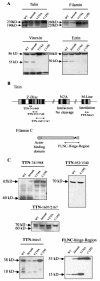
Capn3 cleaves talin, filamins A and C, vinexin, ezrin, and titin Z-disk and M-line domains. (A) Western blot analysis was done on extracts from mouse myoblasts transiently transfected with pTOM-Capn3 (WT), pTOM-Capn3ΔExon6 (Δexon6), pSRD-Capn3Y274A/pEYFP (Y274A), and pSRD-Capn3C129S/pEYFP (C129S). Talin was revealed with a monoclonal antibody. The unprocessed talin form at 230 kDa and a 190-kDa proteolytic fragment are indicated. Filamin A was detected with a monoclonal antibody. The unprocessed filamin A form and a 220-kDa proteolytic fragment are indicated. Vinexin was revealed with a polyclonal antibody. The unprocessed alpha isoform of vinexin at 86 kDa and a proteolytic fragment at 50 kDa are indicated. Ezrin was stained with a polyclonal antibody. The unprocessed ezrin form at 80 kDa and a proteolysis product at 50 kDa are indicated. (B) Organization of titin Z disk, N2A, and M line and filamin C. Gray, black, and white boxes represent repetitive immunoglobulin-like, unique, and Z-repeat regions of titin, respectively. Known relationships between Capn3 and titin are indicated below the corresponding scheme. Cloned fragments are represented by lines with double arrows. The titin domain corresponding to residues 741 to 948 was fused to eGFP. Titin domains corresponding to residues 952 to 1540 and 1607 to 2167 and the Mex5 domain were tagged with the V5 epitope. The 53-kDa COOH terminus of filamin C was tagged with the V5 epitope. (C) Western blot analysis was done on extracts from mouse myoblasts cotransfected with the corresponding plasmids and pSRD-wt-Capn3 (WT), pSRD-Capn3ΔExon6 (Δexon6), pSRD-Capn3Y274A (Y274A), or pSRD-Capn3C129S (C129S) as a negative control. Titin and filamin C domains were revealed with a monoclonal anti-V5 antibody and a polyclonal anti-GFP antibody. The unprocessed form of TTN-741/948 is shown at 65 kDa. A proteolytic 40-kDa fragment was observed for wt Capn3 and Capn3ΔExon6, but not for Capn3Y274A and Capn3C129S transfected cells. TTN-952/1540 shows a single band at 70 kDa with no apparent proteolytic cleavage. Full-length TTN-1607/2167 and a proteolytic product are indicated at 70 and 60 kDa, respectively. The full-length Mex5 domain and a proteolytic fragment are indicated at 38 and 18 kDa, respectively. The full-length filamin C domain and a proteolytic fragment are indicated at 53 and 15 kDa, respectively.
FIG. 4.
FIG. 4.
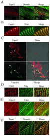
Capn3 colocalizes with its substrates in vivo at the Z disk, M line, costameres, and MTJ. Longitudinal sections of mouse tibialis anterior muscles were stained with anti-Capn3 RP1 or RP4 antibody.Images were visualized by confocal microscopy. White and green scale bars, 2 and 10 μm, respectively. (A) The Z disk was detected with a monoclonal anti-desmin antibody. Red arrows indicate colocalization (yellow) of Capn3 with desmin at the Z disk. (B) The N2A line was detected with the monoclonal anti-titin NCL-TTN antibody. White arrowheads show Capn3 staining at the M line. (C) MTJ were localized with respect to their enrichment in vinculin and to the vicinity of tendons (red arrow). White arrows indicate colocalization (yellow) of Capn3 with vinculin at the MTJ. (D) The N2A line was detected with the monoclonal anti-titin NCL-TTN antibody. (E) Talin was revealed with a monoclonal antibody. White arrowheads show colocalization (yellow) of Capn3 with talin. (F) Ezrin was localized with a specific rabbit polyclonal antibody. White arrowheads indicate colocalization (yellow) of ezrin with desmin at the Z disk.
FIG. 5.
FIG. 5.
(A) Model for Capn3 activation. Capn3 is mainly present in a full-length inactive state in skeletal muscle, presumably through its binding to titin. Upon receiving an activation signal, a subset of Capn3 molecules undergoes intramolecular autolysis in S1. This first event allows the complete autoprocessing of these molecules, consisting of cleavage of S2 and S3. These fully activated Capn3 molecules can thereafter intermolecularly autolyze other Capn3 molecules which have not received the activation signal. This ultimate step generates an amplification cascade leading to global activation of the Capn3 pool. (B) Model for Capn3 cytoskeleton remodeling in skeletal muscle. The main components of costameres are depicted: integrins alpha and beta, talin, the dystrophin glycoprotein complex (DGC), and dystrophin. Examples of components of the Z disk (alpha actinin) and of the M line (M protein and myomesin) are also shown. Inactive Capn3 receives an activation signal, potentially of mechanical origin and occurring at the N2A line. Upon activation, Capn3 is able to cleave titin (Z-disk and Mex5 domains), filamin C, talin, vinexin, ezrin, and possibly other proteins at the Z disk, M line, and costameres. Regarding our knowledge of the function and structure of these proteins, the cleavage products could produce gains of function, inducing an adaptive response of muscle cells to the initial activation signal.

References

    1. Andreoli, C., M. Martin, R. Le Borgne, H. Reggio, and P. Mangeat. 1994. Ezrin has properties to self-associate at the plasma membrane. J. Cell Sci. 107:2509-2521. - PubMed
    1. Baghdiguian, S., M. Martin, I. Richard, F. Pons, C. Astier, N. Bourg, R. T. Hay, R. Chemaly, G. Halaby, J. Loiselet, L. V. Anderson, A. Lopez de Munain, M. Fardeau, P. Mangeat, J. S. Beckmann, and G. Lefranc. 1999. Calpain 3 deficiency is associated with myonuclear apoptosis and profound perturbation of the IkappaB alpha/NF-kappaB pathway in limb-girdle muscular dystrophy type 2A. Nat. Med. 5:503-511. - PubMed
    1. Belkin, A. M., N. I. Zhidkova, F. Balzac, F. Altruda, D. Tomatis, A. Maier, G. Tarone, V. E. Koteliansky, and K. Burridge. 1996. Beta 1D integrin displaces the beta 1A isoform in striated muscles: localization at junctional structures and signaling potential in nonmuscle cells. J. Cell Biol. 132:211-226. - PMC - PubMed
    1. Bialkowska, K., S. Kulkarni, X. Du, D. E. Goll, T. C. Saido, and J. E. Fox. 2000. Evidence that beta3 integrin-induced Rac activation involves the calpain-dependent formation of integrin clusters that are distinct from the focal complexes and focal adhesions that form as Rac and RhoA become active. J. Cell Biol. 151:685-696. - PMC - PubMed
    1. Calderwood, D. A., R. Zent, R. Grant, D. J. Rees, R. O. Hynes, and M. H. Ginsberg. 1999. The talin head domain binds to integrin beta subunit cytoplasmic tails and regulates integrin activation. J. Biol. Chem. 274:28071-28074. - PubMed

Publication types

MeSH terms

LinkOut - more resources