. 2003 Dec;23(24):9251-61.
doi: 10.1128/MCB.23.24.9251-9261.2003.

Ubiquitin depletion as a key mediator of toxicity by translational inhibitors

Affiliations

Ubiquitin depletion as a key mediator of toxicity by translational inhibitors

John Hanna et al. Mol Cell Biol. 2003 Dec.

Abstract

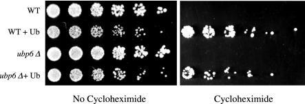
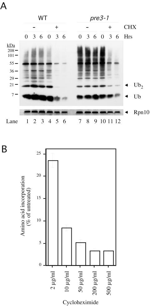
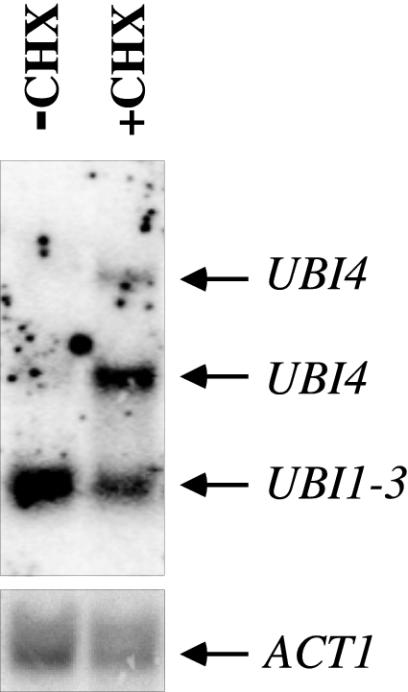
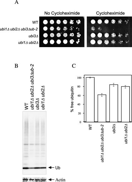
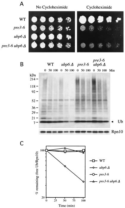
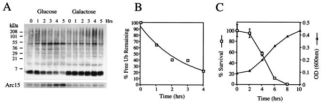
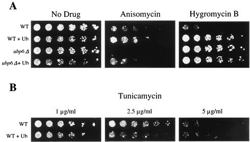
Cycloheximide acts at the large subunit of the ribosome to inhibit translation. Here we report that ubiquitin levels are critical for the survival of Saccharomyces cerevisiae cells in the presence of cycloheximide: ubiquitin overexpression confers resistance to cycloheximide, while a reduced ubiquitin level confers sensitivity. Consistent with these findings, ubiquitin is unstable in yeast (t(1/2) = 2 h) and is rapidly depleted upon cycloheximide treatment. Cycloheximide does not noticeably enhance ubiquitin turnover, but serves principally to block ubiquitin synthesis. Cycloheximide also induces UBI4, the polyubiquitin gene. The cycloheximide-resistant phenotype of ubiquitin overexpressors is also characteristic of partial-loss-of-function proteasome mutants. Ubiquitin is stabilized in these mutants, which may account for their cycloheximide resistance. Previous studies have reported that ubiquitin is destabilized in the absence of Ubp6, a proteasome-associated deubiquitinating enzyme, and that ubp6 mutants are hypersensitive to cycloheximide. Consistent with the model that cycloheximide-treated cells are ubiquitin deficient, the cycloheximide sensitivity of ubp6 mutants can be rescued either by ubiquitin overexpression or by mutations in proteasome subunit genes. These results also show that ubiquitin wasting in ubp6 mutants is proteasome mediated. Ubiquitin overexpression rescued cells from additional translational inhibitors such as anisomycin and hygromycin B, suggesting that ubiquitin depletion may constitute a widespread mechanism for the toxicity of translational inhibitors.


Figures

FIG. 1.
FIG. 1.
Cycloheximide resistance of wild-type and ubp6Δ cells due to ubiquitin overexpression. Threefold serial dilutions of wild-type (WT) cells (SJH30), wild-type cells overexpressing ubiquitin (Ub) (SJH34), ubp6Δ cells (SJH31), and ubp6Δ cells overexpressing ubiquitin (SJH35) were applied as spots to selective plates containing copper sulfate (100 μM) as described with or without 2-μg/ml cycloheximide and grown at 30°C for 3 (left panel) or 7 (right panel) days. Cycloheximide-containing plates were incubated longer to correct for reduced growth rates that result from translational inhibition.
FIG. 2.
FIG. 2.
Depletion of ubiquitin by cycloheximide in wild-type and pre3-1 cells. (A) Wild-type (WT) (YHI29W) or pre3-1 cells (YRG11) were grown in YPD at 30°C in the exponential phase with or without 200-μg/ml cycloheximide (CHX). Aliquots containing equivalent amounts of cells were taken at the indicated time points and analyzed by immunoblotting with antiubiquitin antibody (top panel) or anti-Rpn10 control antibody (bottom panel). Essentially identical results were obtained for the wild-type SUB62 strain (data not shown). (B) Equivalent numbers of wild-type cells pretreated for 1 h with cycloheximide at the indicated concentrations were incubated with 35S-labeled methionine for 15 min. TCA-insoluble material was subjected to scintillation counting. Error bars represent the standard deviation of an experiment carried out in triplicate.
FIG. 3.
FIG. 3.
Induction of the UBI4 gene by cycloheximide. Wild-type (SUB62) cells were grown in the exponential phase for 2 h in the presence (+CHX) or absence (−CHX) of 200-μg/ml cycloheximide. mRNA was isolated and analyzed as described in Materials and Methods. The bands correspond to the UBI1-3 and UBI4 (1.5 and 2.6 kb) genes as indicated (top panel) or ACT1 (bottom panel).
FIG. 4.
FIG. 4.
Cycloheximide sensitivity of a ubi1Δ ubi2Δ ubi3Δub-2 mutant. (A) Threefold serial dilutions of wild-type (WT) (SUB62), ubi1Δ ubi2Δ ubi3Δub-2 (SUB276), ubi3Δ (SUB246), and ubi1Δ ubi2Δ (SUB254) cells were applied as spots to YPD plates with or without 1.0-μg/ml cycloheximide and grown at 30°C for 2 (left panel) or 4 days (right panel). Note that assays of cycloheximide sensitivity are carried out at 0.5 to 1 μg/ml, whereas assays of resistance are carried out at 2 μg/ml, because control cells are sensitive to 2 μg/ml but resistant to 0.5 to 1 μg/ml. (B) Wild-type and mutant cells as described above were grown in YPD at 30°C in the exponential phase. An equivalent number of cells were analyzed for free ubiquitin (Ub) levels by immunoblotting with antiubiquitin antibody followed by 125I-labeled protein A (upper panel). The same immunoblot was stripped and reprobed with antiactin antibody followed by 125I-labeled protein A (lower panel). (C) Quantitation of free ubiquitin levels from the data in panel B. The data shown have been normalized against actin levels to correct for total protein loads. Error bars represent the standard deviations of the experiment carried out in duplicate.
FIG. 5.
FIG. 5.
Ubiquitin wasting in ubp6 mutants is mediated by the proteasome. (A)Threefold serial dilutions of wild-type (WT) (YHI29W), pre3-6 (YRG16), ubp6Δ (SJH42), and pre3-6 ubp6Δ (SJH44) were applied as spots to YPD plates with or without 0.5-μg/ml cycloheximide and grown at 30°C for 2 (left) or 7 days (right). Note that pre3-6 is not more resistant to cycloheximide than is the wild type at this concentration of drug, but only at higher cycloheximide levels. (B) Wild-type (YHI29W), ubp6Δ (SJH42), pre3-6 (YRG16), and pre3-6 ubp6Δ (SJH44) cells were grown in YPD at 30°C in exponential phase. Cycloheximide was added to a final concentration of 50 μg/ml, and aliquots containing equivalent amounts of cells were taken at the indicated time points and analyzed by immunoblotting with an antiubiquitin antibody (upper panel) or anti-Rpn10 control antibody (lower panel). Ub, ubiquitin. (C) Quantitation of the data in panel B. The immunoblot from panel B was stripped and reprobed with the same antiubiquitin antibody or anti-Rpn10 antibody followed by 125I-labeled protein A. The data shown have been normalized against Rpn10 levels, which is necessary to correct for total protein loads. It should be noted that free ubiquitin levels appear to be slightly decreased over the 100-min time course in the wild-type, pre3-6, and pre3-6 ubp6Δ samples (see panel B), but show no decrease when normalized to Rpn10. It is therefore possible that the decay curves of panel C slightly underestimate the rate of disappearance of free ubiquitin.
FIG. 6.
FIG. 6.
Determination of the half-life of ubiquitin. (A) SUB328 cells expressing a single galactose-inducible ubiquitin gene were grown in galactose in exponential phase. At time zero, cells were either transferred to glucose-containing media or maintained on galactose, and aliquots were taken at the indicated time points. For cells growing on glucose, an equal culture volume was taken at each time point to prevent dilution of the total ubiquitin pool by cell division; for cells growing on galactose, an equivalent number of cells were taken. Cells were analyzed by immunoblotting for ubiquitin (upper panel), followed by enhanced chemiluminescence. Loads were normalized by using an antibody to Arc15 as described previously (lower panel). (B) The immunoblot from panel A was stripped and reprobed with the same antiubiquitin antibody, followed by 125I-labeled protein A, and quantitated on a PhosphorImager with NIH Image. There was a lag time of approximately 1 h in ubiquitin (Ub) shutoff by glucose, and therefore the half-life was determined from 1 to 5 h postshift. (C) Cultures of SUB328 growing in glucose or galactose in exponential phase (as in panel A) were sampled at the indicated time point, and an equivalent amount of OD600 units were plated on YPRafGal plates. Colonies were counted 2 days later. The survival curve is expressed as the percent plating efficiency of the glucose-grown culture divided by that of the galactose culture (□). Error bars represent the standard deviation from an experiment carried out in triplicate. Cultures of SUB328 shifted to glucose media were normalized by OD and allowed to grow at 30°C in the exponential phase. ODs were determined at the indicated time points (▵).
FIG. 7.
FIG. 7.
Resistance to multiple translational inhibitors mediated by ubiquitin overexpression. Threefold serial dilutions of wild-type (WT) cells (SJH30), wild-type cells overexpressing ubiquitin (Ub) (SJH34), ubp6Δ (SJH31) cells, and ubp6Δ cells overexpressing ubiquitin (SJH35) were applied as spots to selective plates containing copper sulfate (100 μM) and no drug, anisomycin (50 μg/ml), or hygromycin B (100 μg/ml) (A) or tunicamycin (B) (concentrations as indicated) and grown at 30°C for 2 to 7 days.

Similar articles

Cited by

References

    1. Abraham, A. K., and A. Pihl. 1983. Effect of protein synthesis inhibitors on the fidelity of translation in eukaryotic systems. Biochim. Biophys. Acta 741:197-203. - PubMed
    1. Ausubel, F. M., R. Brent, R. E. Kingston, D. D. Moore, J. G. Seidman, J. A. Smith, and K. Struhl (ed.). 1994. Current protocols in molecular biology. John Wiley and Sons, New York, N.Y.
    1. Brostrom, C. O., and M. A. Brostrom. 1998. Regulation of translational initiation during cellular responses to stress. Prog. Nucleic Acid Res. Mol. Biol. 58:79-125. - PubMed
    1. Burbea, M., L. Dreier, J. S. Dittman, M. E. Grunwald, and J. Kaplan. 2002. Ubiquitin and AP180 regulate the abundance of GLR-1 glutamate receptors at postsynaptic elements in C. elegans. Neuron 35:107-120. - PubMed
    1. Chen, Y., and P. W. Piper. 1995. Consequences of overexpression of ubiquitin in yeast: elevated tolerances of osmostress, ethanol and canavanine, yet reduced tolerances of cadmium, arsenite and paromomycin. Biochim. Biophys. Acta 1268:59-64. - PubMed

Publication types

MeSH terms

LinkOut - more resources