. 2003 Dec;23(24):9178-88.
doi: 10.1128/MCB.23.24.9178-9188.2003.

The replication fork barrier site forms a unique structure with Fob1p and inhibits the replication fork

Affiliations

The replication fork barrier site forms a unique structure with Fob1p and inhibits the replication fork

Takehiko Kobayashi. Mol Cell Biol. 2003 Dec.

Abstract

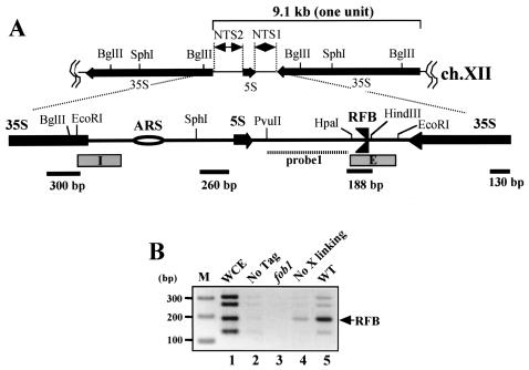
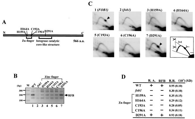
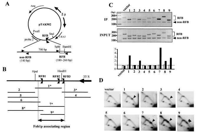
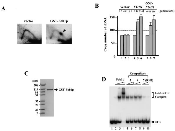
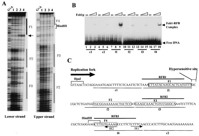
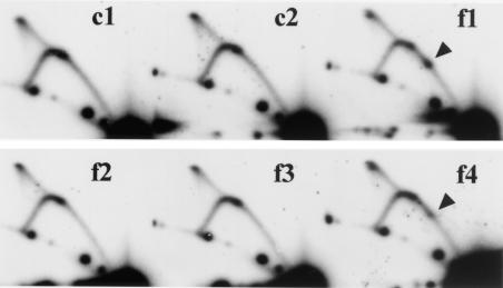
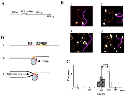
The replication fork barrier site (RFB) is an approximately 100-bp DNA sequence located near the 3' end of the rRNA genes in the yeast Saccharomyces cerevisiae. The gene FOB1 is required for this RFB activity. FOB1 is also necessary for recombination in the ribosomal DNA (rDNA), including increase and decrease of rDNA repeat copy number, production of extrachromosomal rDNA circles, and possibly homogenization of the repeats. Despite the central role that Foblp plays in both replication fork blocking and rDNA recombination, the molecular mechanism by which Fob1p mediates these activities has not been determined. Here, I show by using chromatin immunoprecipitation, gel shift, footprinting, and atomic force microscopy assays that Fob1p directly binds to the RFB. Fob1p binds to two separated sequences in the RFB. A predicted zinc finger motif in Fob1p was shown to be essential for the RFB binding, replication fork blocking, and rDNA recombination activities. The RFB seems to wrap around Fob1p, and this wrapping structure may be important for function in the rDNA repeats.


Figures

FIG. 1.
FIG. 1.
(A) Structure of rDNA repeats in S. cerevisiae. A single unit of rDNA consists of two transcribed genes (5S and 35S RNA genes) (the direction of transcription is indicated by arrows) and two nontranscribed regions (NTS1 and NTS2). The 35S rRNA gene is transcribed by PolI, while the 5S rRNA gene is transcribed by PolIII. The NTS and its surrounding regions are expanded. Two DNA elements related to DNA replication, the origin of replication (ARS) and the RFB, are located in NTS2 and NTS1, respectively. The RFB located near the end of the 35S rRNA gene allows progression of the replication fork in the direction of 35S rRNA transcription but not in the opposite direction (3, 35). Probe 1 (striped bar) is a probe used for Southern hybridization in 2D analysis. The lower bars show locations of fragments amplified by PCR for ChIP assay. The four fragments are located about every 1 kb. The lengths of the PCR products are shown below by bars. E and I (boxed) are elements of HOT1. (B) ChIP assay. The inverse image of PCR products resolved on ethidium bromide-stained 2.6% agarose gels is shown. Fob1-FLAGp was expressed in the fob1 strain (NOY408-1bf), and PCR was performed on chromatin fragments after the immunoprecipitation. Samples were prepared from the whole-cell extract (WCE) before the precipitation (lane 1), from NOY408-1b with Fob1p not tagged (lane 2), and from NOY408-1bf with Fob1-FLAGp not expressed (lane 3), with Fob1-FLAGp expressed but without prior cross-linking before precipitation (lane 4), and with Fob1-FLAGp expressed (lane 5). The position of the RFB fragment is indicated by an arrow. Lane M, 100-bp ladder marker (Invitrogen).
FIG. 2.
FIG. 2.
Effects of various FOB1 mutations. (A) Structure of Fob1p. The gray bar shows the 566-amino-acid (a.a.) Fob1p. Regions corresponding to the predicted zinc finger motif and integrase catalytic core-like structure are shown below the bar. Positions of mutated amino acids are indicated above the bar, and the mutations are labeled according to convention. The N and C termini are indicated. (B) ChIP assay with the mutated Fob1p constructs. Various mutated Fob1-FLAGp were expressed in the fob1 strain (NOY408-1bf), and the associated DNA was precipitated with an anti-FLAG monoclonal antibody (Stratagene) after cross-linking. Lane 1, Fob1-FLAGp was expressed; lane 2, Fob1-FLAGp was not expressed; lane 3 to 7, each mutated Fob1-FLAGp was expressed, as indicated. The position of the RFB fragment is indicated by an arrow. (C) Replication fork-blocking activity at the RFB site in the mutated FOB1strains was analyzed by 2D analysis. DNA was prepared from the strains used for panel B, digested with BglII and SphI, and subjected to 2D analysis followed by Southern hybridization with probe 1 (Fig. 1A). Spots indicated by arrowheads show accumulation of Y-shaped DNA molecules at the RFB site. A diagram of the migration pattern is shown in the lower right panel, with the structures of the replication intermediate molecules shown above the pattern. (D) Summary of the effects of the mutations in FOB1. R.A., RFB, and R.R., RFB-associating activity, replication fork-blocking activity, and recombination rate, respectively. + and −, wild-type level and fob1 level, respectively. The recombination rates were measured by determining the frequency of loss of a URA3 marker inserted in the rDNA repeats. The values are the averages from three independent experiments. Standard deviations (SD) are shown in parentheses.
FIG. 3.
FIG. 3.
Identification of Fob1p association sites in the RFB. (A) Structure of pTAK902.1 to -9, which contain various subfragments of the RFB. RFB subfragments were inserted (between SphI and BamHI sites) near the 2μm replication origin, where they are predicted to inhibit the clockwise-moving replication fork. Positions of PCR primer sets used for the ChIP assay are shown by arrowheads (non-RFB and RFB). (B) Positions of the subfragments which were inserted into the SphI-BamHI site of pTAK902. The RFB1, RFB2, and RFB3 sites and the direction of 35S rDNA transcription are indicated. Numbers with asterisks indicate fragments that were associated with Fob1p (C) and inhibited the replication fork in the plasmid (D). (C) ChIP analysis by Fob1-FLAGp. Samples were prepared from NOY408-1bf strains carrying pTAK901 (Fob1-FLAGp expression vector) and pTAK902.1 to -9. In the top panel, DNA immunoprecipitated (IP) by the anti-FLAG antibody was used as the template, and in the middle panel, total DNA (whole-cell extract) was used as the template. Primer sets used for the ChIP assay are shown in panel A. Non-RFB and RFB primer sets amplified the vector sequence (lower bands) and inserted RFB subfragments (upper bands), respectively. Lane numbers above the panel correspond to the inserted fragments (panel B). vector, no RFB insertion in pTAK902. In bottom panel, the ratios of RFB to non-RFB are plotted. White and black bars indicate the values for input and IP, respectively. The values are relative to that of input vector. (D) Replication fork-blocking activities of various RFB subfragments on plasmid pTAK902. 2D analysis was performed as for Fig. 2C. DNA was prepared from the strains used for panel C, digested with PvuII and StuI, and subjected to 2D analysis followed by Southern hybridization with probe 2 (A). Numbers correspond to those of the subfragments in panel B. Spots indicated by arrowheads show accumulation of Y-shaped DNA molecules at the RFB site. vector, no RFB insertion in pTAK902.
FIG. 4.
FIG. 4.
Binding activity of purified GST-Fob1p. (A) 2D analysis to detect replication fork-blocking activity of GST-Fob1p in vivo. DNA samples were prepared from NOY408-1bf carrying pEG(KT) (vector) or pTAK900 (GST-Fob1p). An arrowhead shows the accumulation of Y-shaped molecules, indicative of replication fork-blocking activity. (B) rDNA amplification activity of GST-Fob1p in vivo. NOY408-1af was transformed with pEG(KT) (vector), Yep-FOB1 (FOB1) or pTAK900 (GST-FOB1). At 44 and 116 generations after the introduction, DNA was isolated and rDNA copy number was determined. Generation 0 corresponds to DNA that was isolated before transformation. (C) Analysis of purified GST-Fob1p by sodium dodecyl sulfate-polyacrylamide gel electrophoresis. GST-Fob1p was purified by using a GST-affinity column and gel filtration from a crude extract of YK9 with pTAK900. The fusion protein was applied to a 10 to 20% polyacrylamide gel and stained with Bio-Safe Coomassie blue (Bio-Rad). (D) Detection of in vitro binding activity of GST-Fob1p to the RFB fragment by gel shift assay. End-labeled RFB fragments (0.16 ng) (Fig. 3B, fragment 7) were mixed with 0 ng (lane 1) 0.25 ng (lane 2), 2.5 ng (lane 3), and 25 ng (lane 4) of GST-Fob1p, and the mixture was applied to a native 5% polyacrylamide gel. Lanes 5 to 10 show competition assays to detect the binding specificity of the RFB fragment. Here, 0.16 ng of end-labeled RFB fragments, 2.5 ng of GST-Fob1p, and one of the three kinds of cold competitor fragments (fragment 5, 4, or 7 [Fig. 3B]) were used in the assay. Fragments 5 and 4 are RFB flanking sequences, and fragment 7 is the RFB itself. Competitors were used at 1.6 ng (lanes 5, 7, and 9) or 3.2 ng (lanes 6, 8, and 10).
FIG. 5.
FIG. 5.
(A) DNase I footprinting assay with purified GST-Fob1p. A 32P-end-labeled RFB fragment (0.1 ng of fragment 7 in Fig. 3B) was mixed with purified GST-Fob1p and digested with DNase I. Samples were analyzed on an 8% (wt/vol) polyacrylamide sequencing gel. Amounts of GST-Fob1p were as follows: lane 1, 0 ng; lane 2, 2.5 ng; lane 3, 25 ng; lane 4, 250 ng. Lane G+A, Maxam-Gilbert sequencing reaction. (B) Detection of in vitro binding activity of GST-Fob1p to small subfragments of the RFB by gel shift assay. The gel shift assay was performed as for Fig. 4D. The positions of the 32P-end-labeled DNA fragments c1 to f4 are shown in panel C. Amounts of GST-Fob1p were as follows: lanes 1, 4, 7, 10, 13, and 16, 0 ng; lanes 2, 5, 8, 11, 14, and 17, 2.5 ng; lanes 3, 6, 9, 12, 15, and 18, 25 ng. (C) Summary of GST-Fob1p binding sites in the RFB. The DNA sequence around the RFB (∼180 bp) is shown. RFB1, RFB2, and RFB3 are indicated by solid lines above the sequence. c1 to f4, used for the gel shift assay in panel B, are shown below the sequence. Fob1p binding sequences, identified by the footprinting assay (A), are shown by boxes. The box (F1) indicates strong binding, and dotted boxes (F2 to F4) indicate weak binding. A DNase I-hypersensitive site next to F1 is indicated. The direction of the replication fork which is inhibited by the RFB is indicated by an arrow. IR1 and IR2 are inverted repeats.
FIG. 6.
FIG. 6.
Replication fork-blocking activity of small RFB subfragments (24 bp). 2D analysis was performed as for Fig. 3D. DNA was prepared from NOY408-1bf with pTAK901 and pTAKc1 to -f4 (which carry the RFB fragments c1 to f4 [Fig. 5C]), digested with PvuII and StuI, and subjected to 2D analysis followed by Southern hybridization with probe 2 (Fig. 3A). Spots indicated by arrowheads show accumulation of Y-shaped DNA molecules at the RFB site.
FIG. 7.
FIG. 7.
Observation of the Fob1-RFB complex by AFM. (A) Structures of RFB fragments used in AFM analysis. The box shows the RFB fragment (fragment 7 from Fig. 3B), and bars indicate non-rDNA sequence. (B) AFM images of the Fob1p-RFB complex in a quick surface plot format. The illustrations drawn as pink lines (DNA) and white circles (Fob1p) are interpretations of the AFM images. (C) Frequency plot (histogram) of the length of the RFB fragment measured from AFM images. White and hatched boxes indicate the numbers of free DNA molecules and Fob1p-RFB complexes, respectively. The mean fragment lengths are indicated below the graph by dotted lines. (D) Models of the Fob1p-RFB complex. a, the RFB fragment without Fob1p. The RFB1 region is shown in red, and RFB2 and RFB3 are shown in yellow. b, the wrapping model. The RFB sequence wraps around Fob1p. Fob1p may be acting as a dimer in this structure. See text for details. c, a model for how the replication fork is inhibited by the RFB wrapping around Fob1p. The fork is inhibited mainly at RFB1.

References

    1. Bedrosian, C. L., and D. Bastia. 1991. Escherichia coli replication terminator protein impedes simian virus 40 (SV40) DNA replication fork movement and SV40 large tumor antigen helicase activity in vitro at a prokaryotic terminus sequence. Proc. Natl. Acad. Sci. USA 88:2618-2622. - PMC - PubMed
    1. Brewer, B. J., and W. L. Fangman. 1987. The localization of replication origins on ARS plasmids in S. cerevisiae. Cell 51:463-471. - PubMed
    1. Brewer, B. J., and W. L. Fangman. 1988. A replication fork barrier at the 3′ end of yeast ribosomal RNA genes. Cell 55:637-643. - PubMed
    1. Brewer, B. J., D. Lockshon, and W. L. Fangman. 1992. The arrest of replication forks in the rDNA of yeast occurs independently of transcription. Cell 71:267-276. - PubMed
    1. Carrigan, C. M., J. A. Haarsma, M. T. Smith, and R. G. Wake. 1987. Sequence features of the replication terminus of the Bacillus subtilis chromosome. Nucleic Acids Res. 15:8501-8509. - PMC - PubMed

Publication types

MeSH terms