

doi: 10.1128/MCB.23.24.9275-9282.2003.
Affinity purification of specific chromatin segments from chromosomal loci in yeast
Affiliations
- PMID: 14645537
- PMCID: PMC309680
- DOI: 10.1128/MCB.23.24.9275-9282.2003
Item in Clipboard
Affinity purification of specific chromatin segments from chromosomal loci in yeast
Mol Cell Biol.
2003 Dec.
Abstract
Single-copy gene and promoter regions have been excised from yeast chromosomes and have been purified as chromatin by conventional and affinity methods. Promoter regions isolated in transcriptionally repressed and activated states maintain their characteristic chromatin structures. Gel filtration analysis establishes the uniformity of the transcriptionally activated state. Activator proteins interact in the manner anticipated from previous studies in vivo. This work opens the way to the direct study of specific gene regions of eukaryotic chromosomes in diverse functional and structural states.
Figures
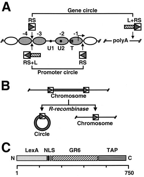
(A) Schematic representation of the genetically modified PHO5 locus. Ovals represent positioned nucleosomes (−1 to −4) on the promoter under repressing conditions. Black dots represent transcription factor binding sites UASP1 (U1) and UASP2 (U2). A square represents the TATA box (T). Arrowheads point to the sites where recognition elements for R recombinase from Z. rouxii (RS) and the LexA operator cluster (L) were inserted into the genomic locus. PHO5 domains released by homologous recombination are indicated. (B) Schematic representation of site-specific homologous recombination. (C) Schematic representation of the recombinant adaptor molecule. LexA, full-length LexA protein from E. coli; NLS, simian virus 40 nuclear localization signal; GR6, Garnier Robson helix 6 from rat plectin; TAP, tandem affinity purification tag. The scale on the bottom reflects the number of amino acids.
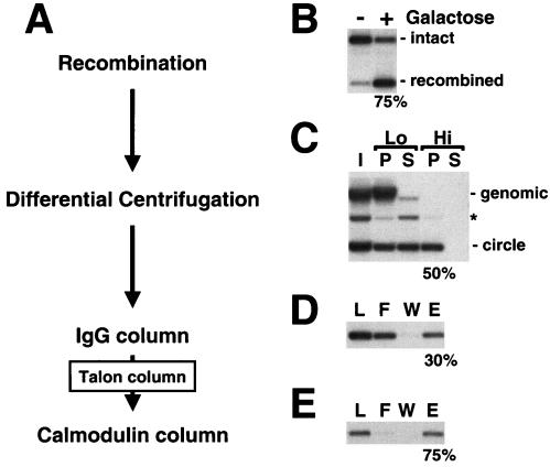
(A) Purification scheme for chromatin circles. (B) Site-specific homologous recombination. The PHO5 locus before (−) and after (+) addition of galactose to induce recombinase expression. Genomic DNA was extracted, digested with ApaI and NciI, subjected to 1% agarose gel electrophoresis, and analyzed by blot hybridization with a probe upstream of the PHO5 locus. Positions of the intact and recombined loci are indicated on the right. (C) Differential centrifugation of yeast whole-cell lysates. I, input; P, pellet; S, supernatant; Lo, low-speed spin; Hi, high-speed spin. DNA was extracted, subjected to 1% agarose gel electrophoresis, and analyzed by blot hybridization with a probe specific for the PHO5 locus. Positions of genomic DNA and circle DNA are indicated on the right. The asterisk marks the position of nicked circle DNA that arose as an artifact during DNA preparation. (D and E) IgG and calmodulin affinity chromatography of chromatin circles. L, load; F, flowthrough; W, wash fraction; E, eluate. DNA was analyzed as described for panel C. Numbers on the bottom of each panel give the percentage of recovery of chromatin circles in each purification step, as determined by quantitative blot hybridization.
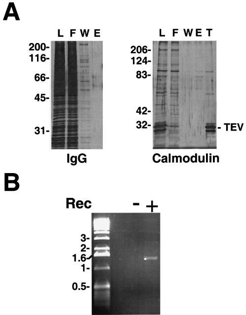
(A) Protein analyses for IgG and calmodulin affinity purification. Samples at different stages of purification were analyzed by SDS-polyacrylamide gel electrophoresis and silver staining. L, F, W, and E are as described in the legend to Fig. 2D and E; T, Talon beads. Numbers on the left of each panel give the positions of molecular size standards (Mr × 10−3). The position of recombinant TEV protease is indicated on the right. (B) DNA analysis of concentrated calmodulin affinity eluates. Calmodulin affinity eluates obtained from 12 liters of yeast culture of strains capable of (+) or deficient in (−) galactose-induced recombinase expression (Rec) were concentrated by centrifugation. DNA was extracted from two thirds of the sample, subjected to 1% agarose gel electrophoresis, and detected by ethidium bromide staining. Numbers on the left give the positions of DNA size standards (in kilobase pairs).
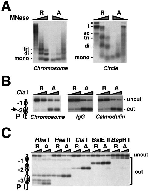
(A) Micrococcal nuclease accessibility of the PHO5 promoter in isolated nuclei and in purified chromatin circles. Nuclei isolated from yeast strains repressed (R) and activated (A) in PHO5 expression were digested with 1, 5, 10, or 20 U of micrococcal nuclease (MNase)/ml for 20 min (Chromosome). PHO5 promoter circles (100 amols of DNA) from the same strains purified by IgG affinity purification were digested with 0.5, 1.5, and 4.5 U of micrococcal nuclease for 5 min (Circle). DNA was extracted, subjected to 2% agarose gel electrophoresis, and analyzed by blot hybridization with a probe spanning nucleosomes N-1 to N-3. The positions of DNA fragments that have been protected by mono-, di-, and trinucleosomes and the positions of supercoiled (sc), linear (l), and nicked (*) DNA are given on the left of each panel. (B and C) Restriction endonuclease accessibility of the PHO5 promoter in isolated nuclei and in purified chromatin circles. Nuclei isolated from yeast strains repressed (R) and activated (A) in PHO5 expression were digested with 50 or 200 U of ClaI restriction endonuclease for 1 h (Chromosome). DNA was extracted, digested with HaeIII, subjected to 1.5% agarose gel electrophoresis, and analyzed by blot hybridization with the probe indicated (P). PHO5 gene circles from the same yeast strains purified by IgG, IgG and calmodulin affinity chromatography (B), or PHO5 promoter circles purified by IgG affinity chromatography (C) were digested with 5 or 20 U of the restriction endonuclease indicated for 30 min. The DNA was extracted, digested with EcoRI (B) or HpaI (C), subjected to 1.5% agarose gel electrophoresis, and analyzed by blot hybridization with the probe indicated. An arrowhead points to the position of the ClaI cutting site in the PHO5 promoter, depicted on the left. Positions of uncut and cut DNA are indicated on the right.
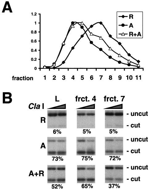
(A) Gel filtration of PHO5 promoter circles. Chromatin circles from yeast strains repressed (R) and activated (A) in PHO5 expression and grown in the absence of the recombinant LexA adaptor molecule were partially purified by differential centrifugation. A TSK-G4000SW column was used to separate promoter circles from the individual preparations and from a 2:1 mixture of activated and repressed promoter circles (A+R). DNA eluting in different fractions (1 to 11) from the column was extracted, subjected to 1% agarose gel electrophoresis, and analyzed by blot hybridization with a probe spanning nucleosomes N-1 to N-3. The DNA concentration relative to that in the respective peak fraction, determined from the radioactivity in the blot, is plotted for each gel filtration experiment on the ordinate. The numbers of the fractions are indicated on the abscissa. (B) Endonuclease accessibility in promoter circles subjected to gel filtration. PHO5 promoter circles present in the load (L) and fractions (frct.) 4 and 7 of the respective gel filtration were digested with ClaI as described in the legend to Fig. 4B. DNA was extracted, digested with HpaI, subjected to 2% agarose gel electrophoresis, and analyzed by blot hybridization with a probe spanning nucleosome N-3. Numbers on the bottom of each panel give the percentage of accessibility as the mean of the plateau values. Positions of uncut and cut DNA are indicated on the right.
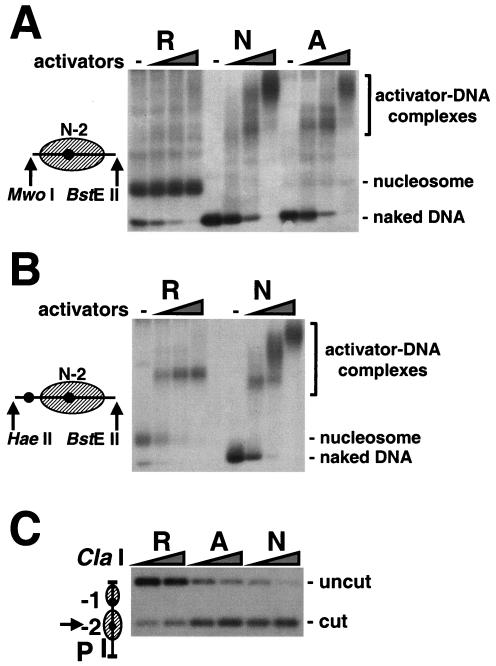
(A and B) Transcription factor binding to natural PHO5 chromatin in vitro. Gene circles purified by IgG affinity chromatography from yeast strains repressed (R) and activated (A) in PHO5 expression and a bacterial plasmid bearing the naked PHO5 promoter sequence (N) were digested with MwoI and BstEII (A) or HaeII and BstEII (B). Restriction digests were incubated for 20 min with or without 30 and 3 nM, 150 and 15 nM, and 750 and 75 nM recombinant Pho4p and Pho2p proteins, respectively. DNA-protein complexes were subjected to native agarose gel electrophoresis and were analyzed by blot hybridization with a probe spanning nucleosome −2. Fragments released upon endonuclease digestion are depicted on the left. Positions of activator-DNA complexes, the nucleosome, and naked DNA are indicated on the right. (C) ClaI accessibility of PHO5 promoter DNA in the presence of transcription factors. Gene circles purified by IgG affinity chromatography from yeast strains repressed and activated in PHO5 expression and a bacterial plasmid bearing the PHO5 promoter sequence were incubated with 750 and 75 nM of recombinant Pho4p and Pho2p proteins, respectively. ClaI accessibility was assessed as described in the legend to Fig. 4B. Positions of uncut and cut DNA are indicated on the right.
References
-
- Ansari, A., T. H. Cheng, and M. R. Gartenberg. 1999. Isolation of selected chromatin fragments from yeast by site-specific recombination in vivo. Methods 17:104-111. - PubMed
-
- Boeger, H., J. Griesenbeck, J. S. Strattan, and R. D. Kornberg. 2003. Nucleosomes unfold completely at a transcriptionally active promoter. Mol. Cell. 11:1587-1598. - PubMed
-
- Godde, J. S., and A. P. Wolffe. 1995. Disruption of reconstituted nucleosomes. The effect of particle concentration, MgCl2 and KCl concentration, the histone tails, and temperature. J. Biol. Chem. 270:27399-27402. - PubMed
Publication types
MeSH terms
Substances
Grants and funding
LinkOut - more resources
Full Text Sources
Other Literature Sources
Molecular Biology Databases