. 2003 Dec;23(24):9293-302.
doi: 10.1128/MCB.23.24.9293-9302.2003.

Regulation of protein tyrosine kinase signaling by substrate degradation during brain development

Affiliations

Regulation of protein tyrosine kinase signaling by substrate degradation during brain development

Lionel Arnaud et al. Mol Cell Biol. 2003 Dec.

Abstract

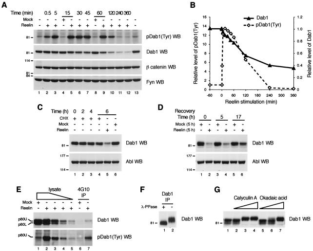
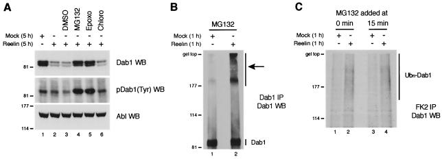
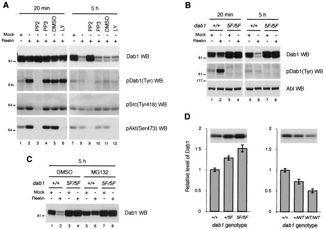
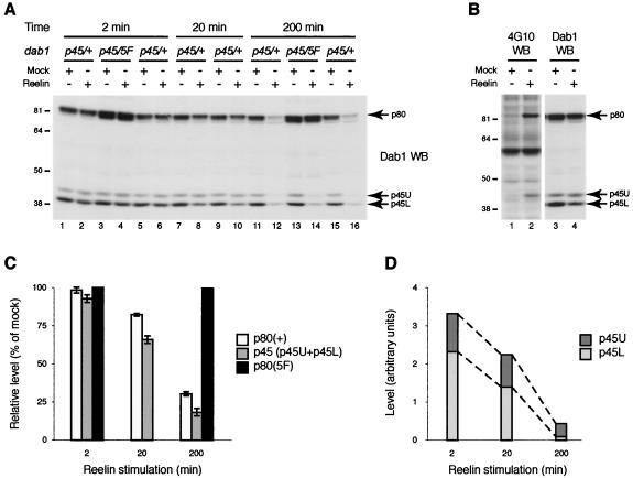
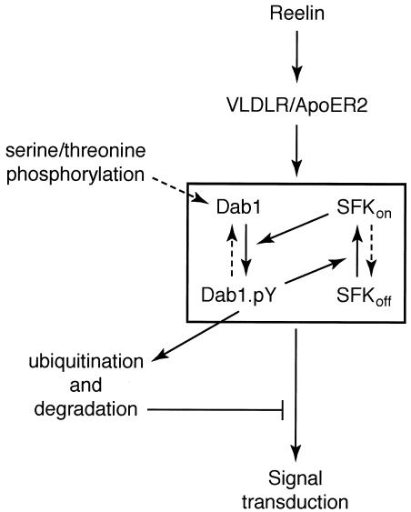
Disabled-1 (Dab1) is a cytoplasmic adaptor protein that regulates neuronal migrations during mammalian brain development. Dab1 function in vivo depends on tyrosine phosphorylation, which is stimulated by extracellular Reelin and requires Src family kinases. Reelin signaling also negatively regulates Dab1 protein levels in vivo, and reduced Dab1 levels may be part of the mechanism that regulates neuronal migration. We have made use of mouse embryo cortical neuron cultures in which Reelin induces Dab1 tyrosine phosphorylation and Src family kinase activation. We have found that Dab1 is normally stable, but in response to Reelin it becomes polyubiquitinated and degraded via the proteasome pathway. We have established that tyrosine phosphorylation of Dab1 is required for its degradation. Dab1 molecules lacking phosphotyrosine are not degraded in neurons in which the Dab1 degradation pathway is active. The requirements for Reelin-induced degradation of Dab1 in vitro correctly predict Dab1 protein levels in vivo in different mutant mice. We also provide evidence that Dab1 serine/threonine phosphorylation may be important for Dab1 tyrosine phosphorylation. Our data provide the first evidence for how Reelin down-regulates Dab1 protein expression in vivo. Dab1 degradation may be important for ensuring a transient Reelin response and may play a role in normal brain development.


Figures

FIG. 1.
FIG. 1.
Reelin stimulation induces degradation of Dab1 in primary cortical neurons. (A) Mouse cortical neuron cultures were left untreated (lanes 1 and 13) or treated with Reelin-containing (lanes 2 and 3, 5 to 7, and 9 to 12) or mock (lanes 4 and 8) supernatant for the indicated times. Total lysates were subjected to SDS-7.5% PAGE and Western blot analysis using an anti-phosphotyrosine antibody to detect tyrosine-phosphorylated Dab1 [pDab1(Tyr) WB], anti-Dab1 antibodies to detect total Dab1 (Dab1 WB), anti-β catenin antibody (β catenin WB), or anti-Fyn antibodies (Fyn WB). (B) Quantification of the data presented in panel A was used to determine relative levels of tyrosine-phosphorylated Dab1 [pDab1(Tyr)] and total Dab1 after Reelin stimulation for various times. In both cases, levels were normalized to those in untreated neuron cultures (set at 1.0). (C) Neuron cultures were treated with 20 μg of cycloheximide/ml (CHX; lanes 1 to 4) and either Reelin-containing (lane 5) or mock (lane 6) supernatant for the indicated times. Total lysates were subjected to SDS-8% PAGE and Western blot analysis using anti-Dab1 antibodies (Dab1 WB) or an anti-Abl antibody (Abl WB) as a loading control. (D) Neuron cultures were treated for 5 h with mock (lanes 1, 3, and 5) or Reelin-containing (lanes 2, 4, and 6) supernatant, washed, and returned to normal growth medium for the indicated times. Total lysates were subjected to SDS-PAGE and Western blot analysis using anti-Dab1 antibodies to detect total Dab1 (Dab1 WB) or an anti-Abl antibody (Abl WB) as a loading control. (E) Dab1 can be resolved into two forms, Dab1 p80L and p80U. Lysates were prepared from neuron cultures treated for 15 min with Reelin-containing or mock supernatant as indicated. A portion was analyzed directly by Western blotting of serial diluted samples: 50 (lanes 1 and 2), 25 (lane 3), 12.5 (lane 4), and 6.25 (lane 5) μg. A second portion was immunoprecipitated (IP) with an anti-phosphotyrosine antibody (4G10 IP), and the immunoprecipitates were analyzed in parallel with the total lysates by SDS-9% PAGE (see Materials and Methods for details) and Western blotting with an anti-phosphotyrosine antibody to detect tyrosine-phosphorylated Dab1 [pDab1(Tyr) WB] or anti-Dab1 antibodies (Dab1 WB). The respective positions of Dab1 p80 of slower electrophoretic mobility (upper band [p80U]) and Dab1 p80 of faster mobility (lower band [p80L]) are indicated. Note that only Dab1 p80U is tyrosine phosphorylated and that Dab1 p80U represents approximately two-thirds of total Dab1 after 15 min of Reelin stimulation. The quantity of Reelin-induced tyrosine-phosphorylated Dab1 in the 4G10 IP is equivalent to that present in 25 μg of total lysate, while the quantity of Dab1 p80U in the 4G10 IP is equivalent to two-thirds of that present in 6.25 μg of total lysate; therefore, after 15 min of Reelin stimulation, tyrosine-phosphorylated Dab1 represents ∼15% (6.25/25 × 2/3 = 1/6 [∼15%]) of total Dab1 equivalent to ∼25% (6.25/25 = 1/4 [25%]) of Dab1 p80U. (F) Brain lysates prepared from E16.5 embryos were subjected to immunoprecipitation with anti-Dab1 antibodies (Dab1 IP). The immunoprecipitate was treated with λ protein phosphatase (λ-PPase) (lane 1) or left untreated (lane 2) before analysis using SDS-9% PAGE and Western blotting with anti-Dab1 antibodies (Dab1 WB) was performed. (G) Neuron cultures were treated for 1 h with 0.4 μM (lanes 2 and 5), 2 μM (lanes 3 and 6), or 10 μM (lanes 4 and 7) calyculin A (lanes 2 to 4), okadaic acid (lanes 5 to 7), or DMSO (lane 1). Total lysates were subjected to SDS-9% PAGE and Western blot analysis using anti-Dab1 antibodies (Dab1 WB).
FIG. 2.
FIG. 2.
Dab1 is polyubiquitinated and degraded via the proteasome pathway in response to the presence of Reelin. (A) Mouse cortical neuron cultures were treated for 5 h with mock (lane 1) or Reelin-containing (lanes 2 to 6) supernatant in the presence or absence of DMSO (lane 3), MG132 ([10 μM]) (lane 4), epoxomicin (Epoxo [10 μM]) (lane 5), or chloroquine (Chloro [10 μM]) (lane 6). Total lysates were subjected to SDS-PAGE and Western blot analysis using anti-Dab1 antibodies to detect total Dab1 (Dab1 WB), an anti-phosphotyrosine antibody to detect tyrosine-phosphorylated Dab1 [pDab1(Tyr) WB], or an anti-Abl antibody (Abl WB) as a loading control. (B) Neuron cultures were treated for 1 h with mock (lane 1) or Reelin-containing (lane 2) supernatant in the presence of MG132 (10 μM) to inhibit degradation of polyubiquitinated proteins by the proteasome. Neuron lysates were then subjected to immunoprecipitation with anti-Dab1 antibodies (Dab1 IP), and the immunoprecipitates were analyzed by SDS-7% PAGE and Western blotting with anti-Dab1 antibodies (Dab1 WB). The respective positions of unmodified Dab1 (Dab1) and high-molecular-mass Dab1 species (arrow) are indicated. (C) Neuron cultures were treated as described for panel B except that MG132 was added after 0 min (lanes 1 and 2) or 15 min (lanes 3 and 4) of mock (lanes 1 and 3) or Reelin (lanes 2 and 4) treatment. Ubiquitinated proteins were immunoprecipitated from neuron lysates with the FK2 antibody (FK2 IP) and analyzed by SDS-7% PAGE and Western blotting with anti-Dab1 antibodies (Dab1 WB). The position of polyubiquitinated Dab1 (Ubn-Dab1) is indicated.
FIG. 3.
FIG. 3.
Reelin-induced degradation of Dab1 correlates with tyrosine phosphorylation of Dab1 and activation of SFKs. (A) Mouse cortical neuron cultures were left untreated (lanes 1 and 2 and lanes 7 and 8) or treated with PP2 (10 μM) (lanes 3 and 9), PP3 (10 μM) (lanes 4 and 10), DMSO (lanes 5 and 11), or LY294002 (LY [50 μM]) (lanes 6 and 12) for 30 min before being stimulated with Reelin-containing or mock supernatant as indicated (in the presence of inhibitors) for 20 min (lanes 1 to 6) or 5 h (lanes 7 to 12). Total lysates were subjected to SDS-PAGE and Western blot analysis using anti-Dab1 antibodies to detect total Dab1 (Dab1 WB), an anti-phosphotyrosine antibody to detect tyrosine-phosphorylated Dab1 [pDab1(Tyr) WB], anti-phospho-Src(Tyr418) antibodies [pSrc(Y418) WB], or anti-phospho-Akt(Ser473) antibodies [pAkt(S473) WB]. (B) Neuron cultures were prepared from littermate embryos derived from dab15F/+ parents and subsequently genotyped. The cultures corresponding to dab1+/+ (lanes 1 and 2 and lanes 5 and 6) or dab15F/5F (lanes 3 and 4 and lanes 7 and 8) embryos were treated with Reelin-containing or mock supernatant as indicated for 20 min (lanes 1 to 4) or 5 h (lanes 5 to 8). Total lysates were subjected to SDS-PAGE and Western blot analysis using anti-Dab1 antibodies to detect total Dab1 (Dab1 WB), an anti-phosphotyrosine antibody to detect tyrosine-phosphorylated Dab1 [pDab1(Tyr) WB] or an anti-Abl antibody (Abl WB) as a loading control. (C) Neuron cultures were prepared as described for panel B. The cultures corresponding to dab1+/+ (lanes 1 and 2 and lanes 5 and 6) or dab15F/5F (lanes 3 and 4 and lanes 7 and 8) embryos were treated with Reelin-containing or mock supernatant (as indicated) for 5 h in the presence of DMSO (lanes 1 to 4) or MG132 (lanes 5 to 8). Total lysates were subjected to SDS-PAGE and Western blot analysis using anti-Dab1 antibodies (Dab1 WB). (D) Dab1 protein levels in dab15F/5F and dab1WT/WT E16.5 brains. Brain lysates were prepared from mouse E16.5 embryos derived from dab15F/+ (left graph [n = 18]) or dab1WT/+ (right graph [n = 18]) parents, and Dab1 protein levels were determined by Western blot analysis using anti-Dab1 antibodies (examples are shown above each graph) and the anti-βIII tubulin antibody TUJ1 for normalization of the loading (data not shown) (standard deviation, 1.8%; n = 36). The relative levels of Dab1 proteins were normalized to the average Dab1 level in wild-type embryos (set to 1.0) (standard deviation, 4.1%; n = 11) and were plotted against the genotypes of embryos as indicated. Error bars represent standard deviations.
FIG. 4.
FIG. 4.
Reelin-induced tyrosine phosphorylation of Dab1 is necessary for its degradation. (A) Cortical neuron cultures were prepared from littermate embryos derived from a cross between a dab15F/+ female and a dab1p45/p45 male and subsequently genotyped. Each culture was treated with Reelin-containing or mock supernatant (as indicated) for 2 (lanes 1 to 6), 20 (lanes 7 to 10), or 200 (lanes 11 to 16) min. Total lysates were subjected to SDS-PAGE and Western blot analysis using anti-Dab1 antibodies (Dab1 WB). The respective positions of full-length Dab1 (p80), Dab1 p45 of slower electrophoretic mobility (upper band [p45U]), and Dab1 p45 of faster mobility (lower band [p45L]) are indicated by arrows. (B) Neuron cultures prepared from a dab1p45/+ embryo were treated with Reelin-containing (lanes 1 and 3) or mock (lanes 2 and 4) supernatant for 20 min. Total lysates were subjected to SDS-PAGE and Western blot analysis using anti-Dab1 antibodies to detect total Dab1 (Dab1 WB) and an anti-phosphotyrosine antibody to detect tyrosine-phosphorylated Dab1 (4G10 WB). The respective positions of full-length Dab1 (p80), Dab1 p45U, and Dab1 p45L are indicated by arrows. (C) Quantification of data presented in panel A. Relative levels of different Dab1 forms, wild-type Dab1 [p80(+)], Dab15F [p80(5F)], and total Dab1p45 [p45 (p45U+p45L)] after Reelin stimulation for 2, 20, or 200 min. The values are presented as percentages of the average level of each form in mock-treated samples. Error bars represent standard deviations. (D) Quantification of data presented in panel A. Relative levels of Dab1 p45U and Dab1 p45L after Reelin stimulation for 2, 20, or 200 min are shown. The values are presented as densitometric units.
FIG. 5.
FIG. 5.
Model for regulating Reelin signaling at the level of Dab1. Reelin binds directly to ApoER2 and VLDLR on the surface of migrating neurons, which stimulates Dab1 tyrosine phosphorylation and SFK activation in a positive-feedback loop, and triggers activation of downstream components of the Reelin-signaling pathway. Reelin-induced tyrosine phosphorylation of Dab1 is a limited process affecting a restricted subpopulation of Dab1 molecules, implying the presence of a limiting factor (represented as a box). Dab1 is primed for tyrosine phosphorylation by one or more serine/threonine phosphorylations (likely on serine) that are Reelin independent. Tyrosine-phosphorylated Dab1 is removed from the system by degradation via the ubiquitin-proteasome pathway, resulting in down-regulation and ultimately termination of Reelin signal transduction.

Similar articles

Cited by

References

    1. Arnaud, L., B. A. Ballif, E. Forster, and J. A. Cooper. 2003. Fyn tyrosine kinase is a critical regulator of disabled-1 during brain development. Curr. Biol. 13:9-17. - PubMed
    1. Asher, G., J. Lotem, L. Sachs, C. Kahana, and Y. Shaul. 2002. Mdm-2 and ubiquitin-independent p53 proteasomal degradation regulated by NQO1. Proc. Natl. Acad. Sci. USA 99:13125-13130. - PMC - PubMed
    1. Ballif, B. A., L. Arnaud, and J. A. Cooper. 2003. Tyrosine phosphorylation of Disabled-1 is essential for Reelin-stimulated activation of Akt and Src family kinases. Brain Res. Mol. Brain Res. 117:152-159. - PubMed
    1. Beffert, U., G. Morfini, H. H. Bock, H. Reyna, S. T. Brady, and J. Herz. 2002. Reelin-mediated signaling locally regulates protein kinase B/Akt and glycogen synthase kinase 3β. J. Biol. Chem. 277:49958-49964. - PubMed
    1. Benhayon, D., S. Magdaleno, and T. Curran. 2003. Binding of purified Reelin to ApoER2 and VLDLR mediates tyrosine phosphorylation of Disabled-1. Brain Res. Mol. Brain Res. 112:33-45. - PubMed

Publication types

MeSH terms

LinkOut - more resources