. 2003 Dec;23(24):9327-37.
doi: 10.1128/MCB.23.24.9327-9337.2003.

A stem structure in fibroblast growth factor receptor 2 transcripts mediates cell-type-specific splicing by approximating intronic control elements

Affiliations

A stem structure in fibroblast growth factor receptor 2 transcripts mediates cell-type-specific splicing by approximating intronic control elements

Andrew P Baraniak et al. Mol Cell Biol. 2003 Dec.

Abstract

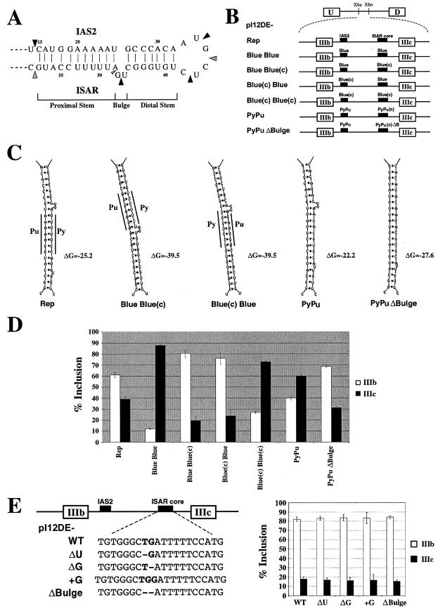
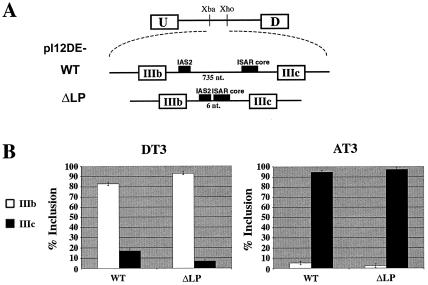
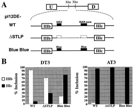
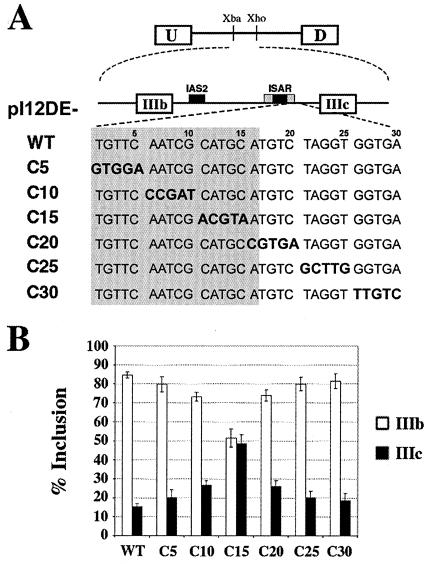
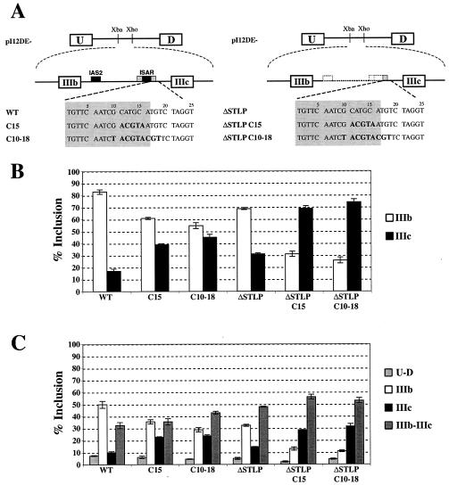
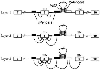
Alternative splicing of fibroblast growth factor receptor 2 (FGFR2) occurs in a cell-type-specific manner with the mutually exclusive use of exon IIIb or exon IIIc. Specific inclusion of exon IIIb is observed in epithelial cells, whereas exon IIIc inclusion is seen in mesenchymal cells. Epithelium-specific activation of exon IIIb and repression of exon IIIc are coordinately regulated by intronic activating sequence 2 (IAS2) and intronic splicing activator and repressor (ISAR) elements in FGFR2 pre-mRNA. Previously, it has been suggested that IAS2 and a 20-nucleotide core sequence of ISAR form a stem structure that allows for the proper regulation of FGFR2 alternative splicing. Replacement of IAS2 and the ISAR core with random sequences capable of stem formation resulted in the proper activation of exon IIIb and repression of exon IIIc in epithelial cells. Given the high degree of phylogenetic conservation of the IAS2-ISAR core structure and the fact that unrelated stem-forming sequences could functionally substitute for IAS2 and ISAR elements, we postulated that the stem structure facilitated the approximation of intronic control elements. Indeed, deletion of the entire stem-loop region and juxtaposition of sequences immediately upstream of IAS2 with sequences immediately downstream of the ISAR core maintained proper cell-type-specific inclusion of exon IIIb. These data demonstrate that IAS2 and the ISAR core are dispensable for the cell-type-specific activation of exon IIIb; thus, the major, if not the sole, role of the IAS2-ISAR stem in exon IIIb activation is to approximate sequences upstream of IAS2 with sequences downstream of the ISAR core. The downstream sequence is very likely a highly conserved GCAUG element, which we show was required for efficient exon IIIb activation.


Figures

FIG. 1.
FIG. 1.
Stem formation dictates exon IIIb inclusion. (A) The structure of an in vitro synthesized 84-nucleotide chimeric RNA molecule containing the rat IAS2 and ISAR core sequences separated by a 6-nucleotide loop was probed with RNase A and RNase T1. Strong RNase A cleavage sites are indicated by large black arrowheads, and strong RNase T1 cleavage sites are indicated by large gray arrowheads. A small gray arrowhead indicates the weak RNase T1 cleavage site. (B) Minigenes used to test the sequence specificity of stem formation for exon IIIb activation in epithelial cells. pI12DE-Rep, pI12DE-Blue Blue(c), pI12DE-Blue(c) Blue, pI12DE-PyPu, and pI12DE-PyPuΔBulge are capable of stem formation, while pI12DE-Blue Blue and pI12DE-Blue(c) Blue(c) are not capable of stem formation. IAS2 and ISAR core or the sequences that replace them are represented as black boxes. (C) Sequence composition of the stems formed by the corresponding minigenes in panel B. Pu and Py demarcate the stretches of purines and pyrimidines in the stem structures. ΔG indicates the predicted Gibbs free energy value for each stem in kcal/mole as calculated by mFold (34). (D) Minigenes that are capable of stem formation recover activation of exon IIIb to various degrees (see Discussion). The percentage of exon inclusion (% IIIb inclusion = 100 × no. of U-IIIb-D transcripts/[no. of U-IIIb-D transcripts + no. of U-IIIc-D transcripts]; % IIIc inclusion = 100 × no. of U-IIIc-D transcripts/[no. of U-IIIb-D transcripts + no. of U-IIIc-D transcripts]) for the minigenes in panel B that were stably transfected into DT3 cells was determined by Invader RNA assay. (E) The two-nucleotide bulge in the stem structure is not necessary for IIIb inclusion. The left panel shows the minigenes used to test the effects of bulge mutations on exon IIIb inclusion. The right panel shows the quantification of RT-PCR analysis of stably transfected minigenes in DT3 cells.
FIG. 2.
FIG. 2.
Loop sequences are not necessary for cell-type specific exon inclusion. (A) Minigenes used to test the importance of loop sequence on cell-type-specific exon inclusion. pI12DE-WT contains 735 nucleotides between IAS2 and ISAR, whereas pI12DE-ΔLP contains 6 nucleotides between IAS2 and ISAR. IAS2 and ISAR core are indicated as black boxes. (B) Quantification of RT-PCR analysis of stably transfected minigenes in DT3 and AT3 cells.
FIG. 3.
FIG. 3.
Approximation of sequences upstream of IAS2 to sequences downstream of ISAR core allows for the proper cell-type-specific exon inclusion. (A) Minigenes used to test the importance of approximating sequences upstream of IAS2 with sequences downstream of ISAR core. pI12DE-WT is described in the legend to Fig. 2A. The pI12DE-ΔSTLP minigene contains a deletion of the entire IAS2-ISAR core stem-loop structure from the 5′ end of IAS2 to the 3′ end of ISAR core, which mimics the predicted outcome of IAS2-ISAR stem formation. The pI12DE-Blue Blue minigene, which is not capable of stem formation, is described in the legend to Fig. 1D. IAS2 and ISAR core are indicated as black boxes. (B) Quantification of RT-PCR analysis of stably transfected minigenes in DT3 and AT3 cells.
FIG. 4.
FIG. 4.
A GCAUG element immediately downstream of ISAR core plays a role in exon IIIb inclusion. (A) Minigenes with five-nucleotide mutations downstream of ISAR core. The five-base-pair substitution mutations are indicated with bold print. IAS2 and ISAR core are represented as black boxes; ISAR core resides within the full ISAR element (represented as a gray box). The nucleotides in the gray box are within ISAR. (B) Quantification of RT-PCR analysis of stably transfected minigenes in DT3 cells.
FIG. 5.
FIG. 5.
A GCAUG element is critical for activation of exon IIIb in minigenes lacking IAS2-ISAR stem structure. (A) Schematic of minigene constructs used in panels B and C. The mutated nucleotides are indicated in bold print. IAS2 and ISAR core are represented as black boxes; ISAR core resides within the full ISAR element (represented as a gray box). The nucleotides in the gray box are within ISAR. (B) The percent inclusion among single-inclusion transcripts (U-IIIb-D and U-IIIc-D) for minigenes in panel A, which were stably transfected into DT3 cells, was determined by Invader RNA assay (e.g., % U-IIIb-D = 100 × no. of U-IIIb-D transcripts/[no. of U-IIIb-D transcripts + no. of U-IIIc-D transcripts]). (C) Quantification of all spliced products for minigenes in panel A, which were stably transfected in DT3 cells, was determined by Invader RNA assay (e.g., % U-IIIb-D = 100 × no. of U-IIIb-D transcripts/[no. of U-D transcripts + no. of U-IIIb-D transcripts + no. of U-IIIc-D transcripts + no. of U-IIIb-IIIc-D transcripts]).
FIG. 6.
FIG. 6.
Layers of control in the alternative splicing of FGFR2 transcripts. The schematic shows three layers of control that determine the choice between inclusion of exon IIIb and exon IIIc. A primary transcript from the FGFR2 gene is shown containing the IAS2-ISAR core structure within intron 8. Layer 1 shows the negative effect of weak splice sites on the definition of exon IIIb. Layer 2 represents the influence of silencer elements that regulate exon IIIb inclusion. These elements operate in both mesenchymal and epithelial cells. Layer 3 depicts epithelial-specific activity that promotes inclusion of IIIb and repression of IIIc. We propose that approximation of sequences downstream of the ISAR core to the vicinity of intronic silencers counters these elements and promotes inclusion of exon IIIb.

Similar articles

Cited by

References

    1. Ahmed, Y. F., G. M. Gilmartin, S. M. Hanly, J. R. Nevins, and W. C. Greene. 1991. The HTLV-I Rex response element mediates a novel form of mRNA polyadenylation. Cell 64:727-737. - PubMed
    1. Blanchette, M., and B. Chabot. 1997. A highly stable duplex structure sequesters the 5′ splice site region of hnRNP A1 alternative exon 7B. RNA 3:405-419. - PMC - PubMed
    1. Bratt, E., and M. Ohman. 2003. Coordination of editing and splicing of glutamate receptor pre-mRNA. RNA 9:309-318. - PMC - PubMed
    1. Brown, P. H., L. S. Tiley, and B. R. Cullen. 1991. Effect of RNA secondary structure on polyadenylation site selection. Genes Dev. 5:1277-1284. - PubMed
    1. Carlo, T., D. A. Sterner, and S. M. Berget. 1996. An intron splicing enhancer containing a G-rich repeat facilitates inclusion of a vertebrate micro-exon. RNA 2:342-353. - PMC - PubMed

Publication types

MeSH terms

Substances