Comparative Study
. 2003 Dec;77(24):12950-60.
doi: 10.1128/jvi.77.24.12950-12960.2003.

Stability and morphology comparisons of self-assembled virus-like particles from wild-type and mutant human hepatitis B virus capsid proteins

Affiliations
Comparative Study

Stability and morphology comparisons of self-assembled virus-like particles from wild-type and mutant human hepatitis B virus capsid proteins

Margaret Newman et al. J Virol. 2003 Dec.

Abstract

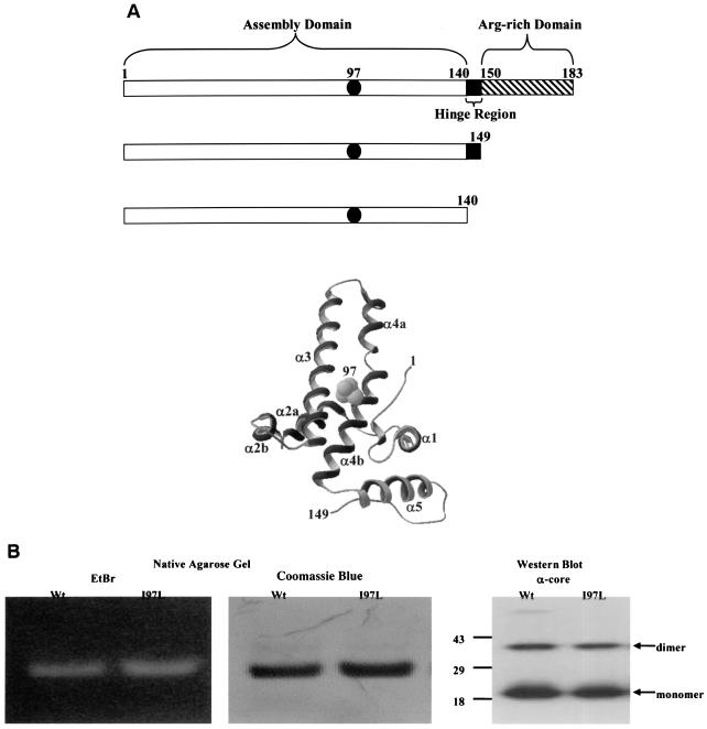
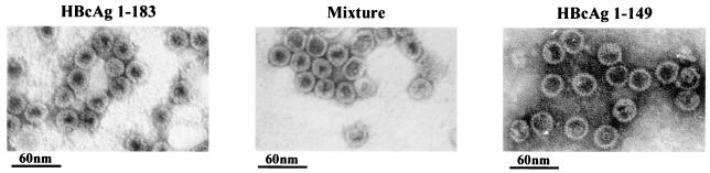
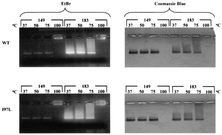
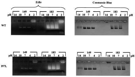
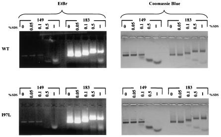
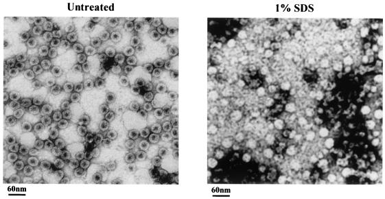
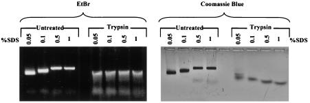
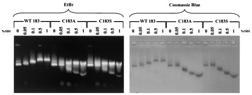
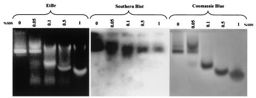
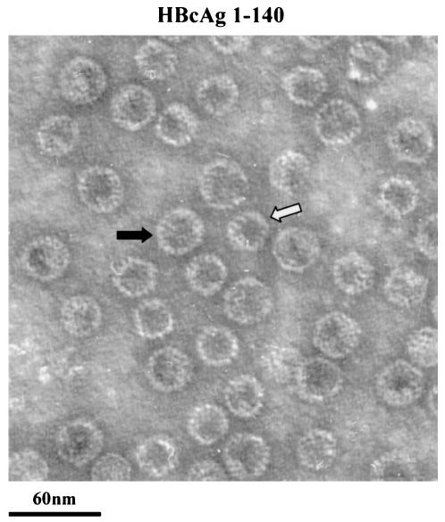
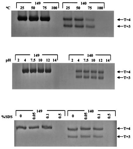
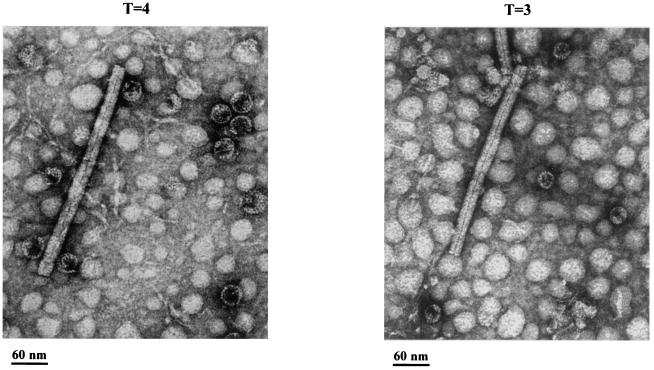
Instead of displaying the wild-type selective export of virions containing mature genomes, human hepatitis B virus (HBV) mutant I97L, changing from an isoleucine to a leucine at amino acid 97 of HBV core antigen (HBcAg), lost the high stringency of selectivity in genome maturity during virion export. To understand the structural basis of this so-called "immature secretion" phenomenon, we compared the stability and morphology of self-assembled capsid particles from the wild-type and mutant I97L HBV, in either full-length (HBcAg1-183) or truncated core protein contexts (HBcAg1-149 and HBcAg1-140). Using negative staining and electron microscopy, full-length particles appear as "thick-walled" spherical particles with little interior space, whereas truncated particles appear as "thin-walled" spherical particles with a much larger inner space. We found no significant differences in capsid stability between wild-type and mutant I97L particles under denaturing pH and temperature in either full-length or truncated core protein contexts. In general, HBV capsid particles (HBcAg1-183, HBcAg1-149, and HBcAg1-140) are very robust but will dissociate at pH 2 or 14, at temperatures higher than 75 degrees C, or in 0.1% sodium dodecyl sulfate (SDS). An unexpected upshift banding pattern of the SDS-treated full-length particles during agarose gel electrophoresis is most likely caused by disulfide bonding of the last cysteine of HBcAg. HBV capsids are known to exist in natural infection as dimorphic T=3 or T=4 icosahedral particles. No difference in the ratio between T=3 (78%) and T=4 particles (20.3%) are found between wild-type HBV and mutant I97L in the context of HBcAg1-140. In addition, we found no difference in capsid stability between T=3 and T=4 particles successfully separated by using a novel agarose gel electrophoresis procedure.


Figures

FIG. 1.
FIG. 1.
(A) Functional domains and three-dimensional structure of HBV core protein. Filled circles represent a wild-type isoleucine or a mutant leucine at amino acid 97. Truncated wild-type and mutant I97L clones consist of either amino acids 1 to 140 or amino acids 1 to 149 lacking the arginine-rich carboxy termini (▧). A hinge region, which connects the capsid assembly and arginine-rich domains, is exposed on the capsid surface and is protease sensitive (24). The ribbon diagram depicts the three-dimensional structure of the core monomer (3, 6, 32) and the location of amino acid 97. A Swiss PDB Viewer was used to render the diagram from 1QGT obtained from GenBank. (B) Characterizations of the recombinant HBcAg1-149 particles by native agarose gel electrophoresis and immunoblot analysis. Encapsidated nucleic acids were stained with EtBr, and the same gel was subsequently stained with protein-specific Coomassie blue. A rabbit polyclonal anticore antibody was used to detect the HBcAg (27).
FIG. 2.
FIG. 2.
Electron micrographs of full-length (HBcAg1-183, left panel) and truncated (HBcAg1-149, right panel) HBcAg capsids. Capsids were negatively stained with 2% uranyl acetate and photographed by using a Philips EM201 electron microscope. The middle panel is a 1:1 mixture of full-length and truncated HBcAg capsids. HBcAg1-183 has a thick-walled appearance, whereas the HBcAg1-149 has a thin-walled appearance with more interior space.
FIG. 3.
FIG. 3.
Similar stabilities of HBV capsids were observed at various temperatures irrespective of the capsid origins (wild type [top panels] or mutant [lower panels]) or size (full-length, HBcAg1-183 or truncated HBcAg1-149). A total of 20 μg of capsid protein was incubated for 15 min at the indicated temperatures before electrophoresis in a 1% agarose gel containing 0.5 μg of EtBr/ml (left). The same gels were subsequently stained with Coomassie blue (right).
FIG. 4.
FIG. 4.
Effects of pH on capsid stability are apparent at pH 2 and pH 14. Portions (20 μg) of E. coli-expressed capsid preparations were incubated in TBS buffers at various pH’s for 30 min at 37°C before electrophoresis in a 1% agarose gel. The upper panels show the results of EtBr (left panel) and Coomassie blue (right panel) staining of the wild-type capsid preparations containing full-length (HBcAg1-183) or truncated (HBcAg1-149) core proteins. The results of the pH challenges with the mutant I97L capsids are shown in the lower panels.
FIG. 5.
FIG. 5.
Stability of E. coli-expressed capsids in the presence of SDS. SDS was added to 20-μg portions of capsid preparations to reach the indicated concentrations. The mixtures were immediately run on a 1% agarose gel containing 0.5 μg of EtBr/ml (left panels) and stained with Coomassie blue (right panels). Wild-type (top panels) and mutant I97L (bottom panels) core proteins, either truncated (HBcAg1-149) or full length (HBcAg1-183), were tested. Note that the truncated capsid particles exhibit a downshift pattern at 0.5 and 1% SDS, whereas the full-length HBcAg1-183 particles exhibit an upshift.
FIG. 6.
FIG. 6.
Electron micrographs of full-length HBcAg1-183 particles with or without SDS treatment. Capsids consisting of wild-type full-length core proteins were negatively stained with 2% uranyl acetate and photographed by using a Philips EM201 electron microscope. The left panel shows typical full-length HBcAg capsids that were not exposed to SDS, and the right panel is from full-length capsid samples treated with 1% SDS. No structured entity can be found in SDS-treated HBcAg1-149 particles (data not shown).
FIG. 7.
FIG. 7.
Trypsin predigestion of HBV full-length capsids eliminated the upshift pattern induced by SDS treatment. Portions (20 μg) of capsids consisting of full-length (HBcAg1-183) core proteins were incubated with 25 U of trypsin at 37°C for 30 min. SDS was then added to full-length capsid preparations, with or without trypsin predigestion, to reach the desired concentrations as indicated. They were then run on a 1% agarose gel containing 0.5 μg of EtBr/ml (left panel) and stained with Coomassie blue (right panel). Note that in the left panel, trypsin-digested samples exhibited a slight upshift banding pattern. Further characterizations of this slight upshift banding are shown in Fig. 9.
FIG. 8.
FIG. 8.
Loss of the SDS-induced upshift pattern of E. coli-expressed C183A and C183S mutant capsids. Portions (20 μg) of capsid preparations of wild-type and mutant C183A or C183S were incubated with the indicated concentrations of SDS. The mixtures were immediately run on a 1% agarose gel containing 0.5 μg of EtBr/ml (left panels) and stained with Coomassie blue (right panels). Note that in the right panel the mutant capsid particles exhibit a downshift pattern at 0.1, 0.5, and 1% SDS, whereas wild-type 183 (WT 183) exhibits an upshift. In the left panel, in addition to the downshift banding pattern, there are strong EtBr signals slightly upshifted after 0.1% SDS in samples C183A and C183S. Unlike the continuous upshift pattern in wild-type HBV, the upshift banding pattern in mutants C183A and C183S did not continue to upshift from 0.1% to 1% SDS. The very low-molecular-weight faint signals in the left panel, stained by EtBr in samples treated with 0.05 to 1% SDS, are likely to be small RNA species of E. coli origin.
FIG. 9.
FIG. 9.
Identification of HBV-specific nucleic acids released from mutant C183S capsid particles after SDS treatment. SDS was added to 20 μg of HBcAg C183S capsid preparations to reach the indicated concentrations. The mixtures were immediately run on a 1% agarose gel containing 0.5 μg of EtBr/ml (left panel) and blotted onto nitrocellulose, and the gel was subsequently stained with Coomassie blue (right panel). The center panel is a Southern blot with an HBV adr probe which demonstrated that the packaged nucleic acids are of HBV origin. The faint lower-molecular-weight signals in the left panel, stained by EtBr in samples treated with 0.05, 0.1, and 0.5% SDS, are likely to be small RNA species of E. coli origin.
FIG. 10.
FIG. 10.
Electron micrographs of mutant I97L HBcAg1-140 capsid particles. The proportion of T=3 particles increased to ca. 78% and T=4 particles decreased to ca. 20.3%, when the morphogenic linker peptide 141-149 was deleted (data not shown). Capsids were negatively stained with 2% uranyl acetate and photographed by using a Philips EM201 electron microscope. White arrow, T=3; black arrow, T=4. Similar results were obtained with wild-type HBcAg1-140 particles (data not shown).
FIG. 11.
FIG. 11.
No significant difference in stabilities between T=3 and T=4 wild-type HBcAg1-140 capsid particles separated by GTG low-melting-point agarose gel electrophoresis. Portions (20 μg) of truncated wild-type HBcAg1-149 or HBcAg1-140 particles were treated with different SDS concentrations, pHs, and temperatures. Similar results were obtained with mutant I97L HBcAg1-140 particles (data not shown). Note that, at 75°C, HBcAg1-149 particles seemed to be more stable than HBcAg1-140.
FIG. 12.
FIG. 12.
Electron micrographs of HBcAg1-140 particles prepared by electroelution from the upper band (A) and the lower band (B) on the GTG gel in Fig. 11. The average diameter of the capsid particles in panel A is ∼27 nm (T=4), whereas in panel B it is ∼25 nm (T=3) (38). The rod-like particles are tobacco mosaic virions and were included as an internal standard. The high background in these micrographs is in part due to the impurities coeluted from the GTG gel.

Similar articles

Cited by

References

    1. Beames, B., and R. E. Lanford. 1993. Carboxy-terminal truncations of the HBV core protein affect capsid formation and the apparent size of encapsidated HBV RNA. Virology 194:597-607. - PubMed
    1. Birnbaum, F., and M. Nassal. 1990. Hepatitis B virus nucleocapsid assembly: primary structure requirements in the core protein. J. Virol. 64:3319-3330. - PMC - PubMed
    1. Bottcher, B., S. A. Wynne, and R. A. Crowther. 1997. Determination of the fold of the core protein of hepatitis B virus by electron cryomicroscopy. Nature 386:88-91. - PubMed
    1. Chua, P. K., Y. M. Wen, and C. Shih. 2003. Coexistence of two distinct secretion mutations (P5T and I97L) in hepatitis B virus core produces a wild-type pattern of secretion. J. Virol. 77:7673-7676. - PMC - PubMed
    1. Cohen, B. J., and J. E. Richmond. 1982. Electron microscopy of hepatitis B core antigen synthesized in Escherichia coli. Nature 296:677-679. - PubMed

Publication types

LinkOut - more resources