. 2003 Dec;77(24):12961-7.
doi: 10.1128/jvi.77.24.12961-12967.2003.

Further evidence that papillomavirus capsids exist in two distinct conformations

Affiliations

Further evidence that papillomavirus capsids exist in two distinct conformations

Hans-Christoph Selinka et al. J Virol. 2003 Dec.

Abstract

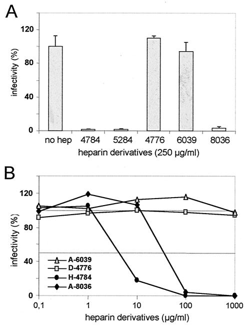
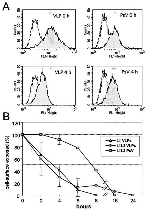
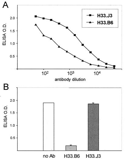
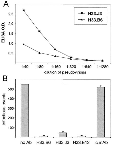
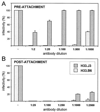
Cell surface heparan sulfate proteoglycans (HSPGs) serve as primary attachment receptors for human papillomaviruses (HPVs). To demonstrate that a biologically functional HPV-receptor interaction is restricted to a specific subset of HSPGs, we first explored the role of HSPG glucosaminoglycan side chain modifications. We demonstrate that HSPG O sulfation is essential for HPV binding and infection, whereas de-N-sulfated heparin interfered with VLP binding but not with HPV pseudoinfection. This points to differences in VLP-HSPG and pseudovirion-HSPG interactions. Interestingly, internalization kinetics of VLPs and pseudovirions, as measured by fluorescence-activated cell sorting analysis, also differ significantly with approximate half times of 3.5 and 7.5 h, respectively. These data suggest that differences in HSPG binding significantly influence postbinding events. We also present evidence that pseudovirions undergo a conformational change after cell attachment. A monoclonal antibody (H33.J3), which displays negligible effectiveness in preattachment neutralization assays, efficiently neutralizes cell-bound virions. However, no difference in H33.J3 binding to pseudovirions and VLPs was observed in enzyme-linked immunosorbent assay and virus capture assays. In contrast to antibody H33.B6, which displays equal efficiencies in pre- and postattachment neutralization assays, H33.J3 does not block VLP binding to heparin, demonstrating that it interferes with steps subsequent to virus binding. Our data strongly suggest that H33.J3 recognizes a conformation-dependent epitope in capsid protein L1, which undergoes a structural change after cell attachment.


Figures

FIG. 1.
FIG. 1.
VLP interaction with HSPGs. (A) VLPs were bound to heparin-BSA-coated microtiter plates in the presence of soluble heparins of different molecular masses (H-4784 [16 kDa], H-5284 [6 kDa], and H-3400 [3 kDa]) or de-N-sulfated (D-4776), de-O-sulfated/acetylated (A-6039), or acetylated heparin (A-8036). Bound VLPs were determined. Binding in the absence of glucosaminoglycan was set as 100%. (B) VLP binding to COS7 cells in the presence of heparin derivatives. Bound VLPs were monitored by Western blot with L1-specific antibody 33L1-7.
FIG. 2.
FIG. 2.
Pseudoinfection in the presence of heparin derivatives. COS7 cells were incubated with pseudovirions in the presence of heparin derivatives at the indicated concentrations. Infectious events were monitored for 72 h postinfection. The number of green fluorescent cells in the absence of inhibitors was set as 100%. Mean values of three independent experiments are shown. (A) Effect of heparin derivatives (H-4776, A-6039, and A-8036) compared to unmodified heparins (H-4784 and H-5284) of various molecular weights. (B) Concentration dependence of the inhibitory effect of heparin derivatives. A value of 100% infectivity corresponds to 370 infected cells.
FIG. 3.
FIG. 3.
Fluorescence-activated cell sorting analysis of VLP and pseudovirus (PsV) internalization. (A) Histograms of cell surface-exposed particles after binding at 4°C (0 h) and further incubation at 37°C (4 h). (B) Time course of internalization of L1 VLPs, L1L2 VLPs, and L1L2 pseudovirions. Mean peak values of seven experiments (± the standard deviations) are shown. The half time of VLP and pseudovirus internalization differs by several hours.
FIG. 4.
FIG. 4.
Interference of monoclonal antibodies with VLP binding to heparin. (A) Titration of two HPV33 VLP-specific monoclonal antibodies (H33.J3 and H33.B6) with HPV33 L1L2 VLPs bound to heparin-BSA-coated microtiter plates. (B) VLPs were incubated with monoclonal antibodies H33.B6 and H33.J3 prior to addition to heparin-BSA-coated plates. The amounts of bound VLPs were determined by using horseradish peroxidase-labeled antibodies. Antibody H33.B6 but not H33.J3 interferes with binding to glucosaminoglycans.
FIG. 5.
FIG. 5.
Reactivity of monoclonal antibodies with HPV33 L1L2 pseudovirions. (A) Interaction of H33.J3 and H33.B6 with heparin-bound pseudovirions. (B) Interaction of monoclonal antibodies with pseudovirions in solution. Pseudovirions were incubated with magnetic beads loaded with the indicated monoclonal antibody. After removal of beads, supernatants were tested for residual infectivity. All HPV33-specific conformation-dependent monoclonal antibodies, but not a control monoclonal antibody (c.mAb), efficiently depleted pseudovirions.
FIG. 6.
FIG. 6.
Pre- and postattachment neutralization assays. (A) For preattachment neutralization, pseudovirions were incubated with the indicated dilutions of monoclonal antibodies for 1 h at 4°C and subsequently added to COS7 cells. Infection was monitored for 72 h after the pseudovirions were replaced by supplemented DMEM. (B) For postattachment neutralization, pseudovirions were bound to COS7 cells for 1 h at 4°C, and cell-bound virions were exposed to antibodies at the indicated dilutions. Antibodies were removed after 1 h at 37°C. A value of 100% infectivity corresponds to 72 infected cells. Antibody H33.J3, but not antibody H33.B6, displays different capacities in pre- and postattachment neutralization assays.

Similar articles

Cited by

References

    1. Baker, T. S., W. W. Newcomb, N. H. Olson, L. M. Cowsert, C. Olson, and J. C. Brown. 1991. Structures of bovine and human papillomaviruses: analysis by cryoelectron microscopy and three-dimensional image reconstruction. Biophys. J. 60:1445-1456. - PMC - PubMed
    1. Bergsdorf, C., C. Beyer, V. Umansky, M. Werr, and M. Sapp. 2003. Highly efficient transport of carboxyfluorescein diacetate succinimidyl ester into COS7 cells using human papillomavirus-like particles. FEBS Lett. 536:120-124. - PubMed
    1. Bernfield, M., M. Götte, P. W. Park, O. Reizes, M. L. Fitzgerald, J. Lincecum, and M. Zako. 1999. Functions of cell surface heparan sulfate proteoglycans. Annu. Rev. Biochem. 68:729-777. - PubMed
    1. Binley, J. M., C. S. Cayanan, C. Wiley, N. Schulke, W. C. Olson, and D. R. Burton. 2003. Redox-triggered infection by disulfide-shackled human immunodeficiency virus type 1 pseudovirions. J. Virol. 77:5678-5684. - PMC - PubMed
    1. Booy, F. P., R. B. Roden, H. L. Greenstone, J. T. Schiller, and B. L. Trus. 1998. Two antibodies that neutralize papillomavirus by different mechanisms show distinct binding patterns at 13 Å resolution. J. Mol. Biol. 281:95-106. - PubMed

Publication types

MeSH terms

LinkOut - more resources