. 2003 Dec;77(24):13005-16.
doi: 10.1128/jvi.77.24.13005-13016.2003.

Changes in small intestinal homeostasis, morphology, and gene expression during rotavirus infection of infant mice

Affiliations

Changes in small intestinal homeostasis, morphology, and gene expression during rotavirus infection of infant mice

Jos A Boshuizen et al. J Virol. 2003 Dec.

Abstract

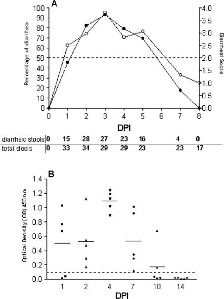
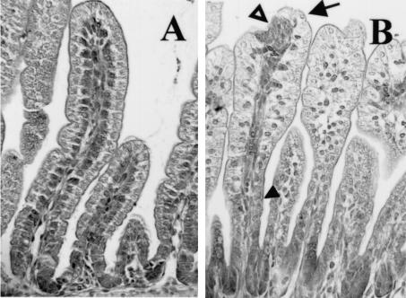
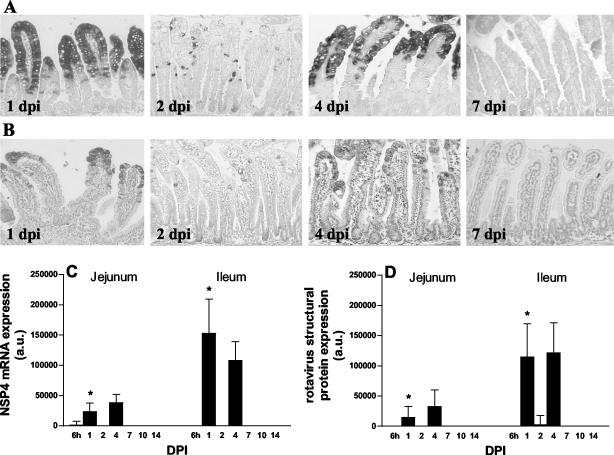
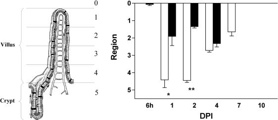
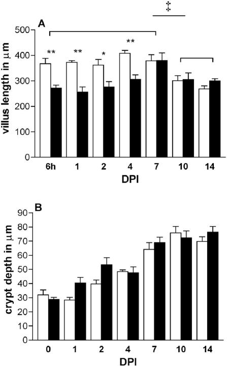
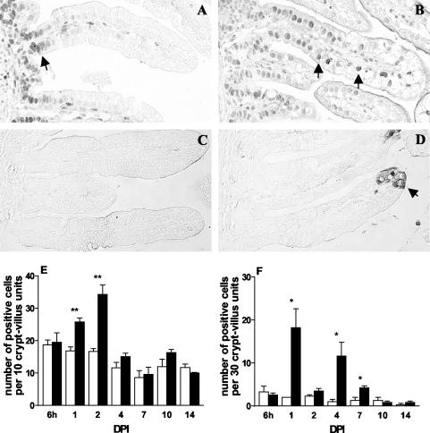
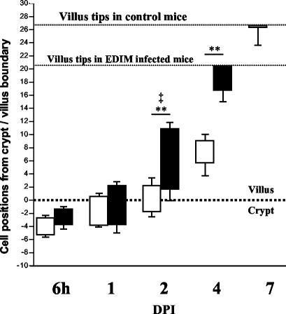
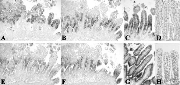
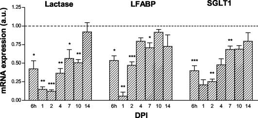
Rotavirus is the most important cause of infantile gastroenteritis. Since in vivo mucosal responses to a rotavirus infection thus far have not been extensively studied, we related viral replication in the murine small intestine to alterations in mucosal structure, epithelial cell homeostasis, cellular kinetics, and differentiation. Seven-day-old suckling BALB/c mice were inoculated with 2 x 10(4) focus-forming units of murine rotavirus and were compared to mock-infected controls. Diarrheal illness and viral shedding were recorded, and small intestinal tissue was evaluated for rotavirus (NSP4 and structural proteins)- and enterocyte-specific (lactase, SGLT1, and L-FABP) mRNA and protein expression. Morphology, apoptosis, proliferation, and migration were evaluated (immuno)histochemically. Diarrhea was observed from days 1 to 5 postinfection, and viral shedding was observed from days 1 to 10. Two peaks of rotavirus replication were observed at 1 and 4 days postinfection. Histological changes were characterized by the accumulation of vacuolated enterocytes. Strikingly, the number of vacuolated cells exceeded the number of cells in which viral replication was detectable. Apoptosis and proliferation were increased from days 1 to 7, resulting in villous atrophy. Epithelial cell turnover was significantly higher (<4 days) than that observed in controls (7 days). Since epithelial renewal occurred within 4 days, the second peak of viral replication was most likely caused by infection of newly synthesized cells. Expression of enterocyte-specific genes was downregulated in infected cells at mRNA and protein levels starting as early as 6 h after infection. In conclusion, we show for the first time that rotavirus infection induces apoptosis in vivo, an increase in epithelial cell turnover, and a shutoff of gene expression in enterocytes showing viral replication. The shutoff of enterocyte-specific gene expression, together with the loss of mature enterocytes through apoptosis and the replacement of these cells by less differentiated dividing cells, likely leads to a defective absorptive function of the intestinal epithelium, which contributes to rotavirus pathogenesis.


Figures

FIG. 1.
FIG. 1.
Percentage of diarrhea (•) and mean diarrheal score (○) (A) and viral antigen shedding (B) in neonatal mice inoculated with rotavirus. Percentage of diarrhea per day was calculated by dividing the number of diarrheic samples by the number of total samples collected each day. A score of ≥2 was considered diarrhea, whereas a score of <2 was considered normal. The mean diarrheal score was determined by dividing the sum of all diarrhea or not-diarrhea scores (1 to 4) by the number of total samples scored each day (numbers are listed at the bottom of the figure). Fecal samples (n = 5 per day) from 0 to 14 dpi were assayed for rotavirus antigen shedding by ELISA (B). Data are expressed as net and mean OD450 and represent individual values obtained for fecal samples. Readings of ≥0.1 are considered positive.
FIG. 2.
FIG. 2.
Histopathological lesions in mouse small intestine (jejunum) during rotavirus infection at 1 dpi. Sections were stained with hematoxylin and eosin. (A) In control animals, enterocytes were clearly polarized and the nuclei were localized at the base of the enterocytes. (B) In infected mice at 1 dpi, histopathological changes were characterized by vacuolization of the enterocytes, swelling of the villus tips (arrow), constriction of the bases, and nuclei that were irregularly positioned within the cells (solid arrowhead). In many villi, lesions seemed to be present at the tips (open arrowhead). Magnification, ×250.
FIG. 3.
FIG. 3.
Kinetics of rotavirus replication in the mouse small intestine. Levels of NSP4 mRNA (A) and protein (B) expression in jejunum at several days postinfection were determined by in situ hybridization and immunohistochemistry, respectively. No rotavirus antigen was detected beyond 7 dpi in any part of the small intestinal epithelium. Quantitative expression of NSP4 mRNA and structural rotavirus proteins were analyzed by RNA and protein dot blotting, respectively (C and D). At 1 dpi, the levels of NSP4 mRNA and structural protein expression were significantly higher in the ileum than in the jejunum. *, P < 0.05 (Student's t test). Magnification, ×150. Error bars, SEM; a.u., arbitrary units.
FIG. 4.
FIG. 4.
The pattern of epithelial vacuolization is more extensive than the pattern of replicating virus. Crypt-villus units in the jejunum were divided into five regions of equal length (see the left part of the figure). The position of the vacuolated cells (open bars) and virus-containing cells (solid bars) on the crypt-villus axis was scored from 1 to 5, representing the regions from the tips of the villi to the crypts. At 1 and 2 dpi, infected cells were exclusively found in the upper part of the villi, whereas vacuolated cells were observed along the entire villi and even at the base of the villi. Values are means + SEM (error bars). * and **, P < 0.01 and P < 0.001, respectively (Student's t test). At the left, a crypt-villus unit is shown and the different regions are indicated.
FIG. 5.
FIG. 5.
Villus length (A) and crypt depth (B) in the jejunum of control mice (open bars) and during rotavirus infection (solid bars). Data from three to five animals at each time point are expressed as mean villus height and mean crypt depth + SEM (error bars). * and **, P < 0.05 and P < 0.01 (Student's t test). Controls at 6 and 7 dpi were compared to controls at 10 and 14 dpi and analyzed by analysis of variance followed by an unpaired t test (‡, P < 0.05).
FIG. 6.
FIG. 6.
Proliferation and apoptosis in jejunum during rotavirus infection as analyzed by staining of PCNA and cleaved caspase-3, respectively. (A) In control animals, proliferating cells were exclusively found in the crypt compartment (arrow). (B) In infected animals at 1 dpi, proliferating cells were observed on up to two-thirds of the length of the villi (arrows). (E) The numbers of proliferating cells in infected animals were strongly increased at 1 and 2 dpi (solid bars) and were comparable to control numbers (open bars) between 4 and 14 dpi (E). (C and F) Apoptotic cells were rarely observed in control animals (open bars). During infection at 1 dpi, however, apoptotic cells were abundantly observed at the villus tips (D) (arrow), and the numbers of apoptotic cells were significantly increased at 1, 4, and 7 dpi (F) (solid bars). * and **, P < 0.05 and P < 0.01 (Student's t test). Error bars, SEM. Original magnification, ×250.
FIG. 7.
FIG. 7.
Cell migration kinetics in the mouse small intestine (ileum) during rotavirus infection. Just before inoculation, mice were injected with BrdU. The positions of the foremost as well as least-progressed BrdU-labeled cells in each crypt-villus unit are expressed as the number of cell positions from the crypt-villus boundary (shown as a dotted line at 0 on the graph). At 6 hpi, BrdU-positive cells in control (open bars) and infected (closed bars) animals were restricted to the crypt compartment. From 2 to 7 dpi, BrdU-positive cells in infected animals migrated significantly higher up the villi than in respective controls (*, P < 0.05; **, P < 0.01) The number of cell positions between the foremost and least-advanced cells was also increased at 2 dpi (‡, P < 0.05). However, in infected animals, BrdU-labeled cells were predominantly lost from the villi at 4 dpi.
FIG. 8.
FIG. 8.
Histological analysis of enterocyte gene expression during rotavirus infection. NSP4 (A), L-FABP (B), beta-actin (E), and SGLT1 (F) mRNA expression in serial small intestinal (jejunum) sections during rotavirus infection as examined by in situ hybridization (at 1 dpi). During massive expression of NSP4 in enterocytes in the upper halves of the villi at 1 dpi (A), enterocyte-specific L-FABP, beta-actin, and SGLT1 mRNA expression was downregulated in these cells (B, E, and F). In control animals, L-FABP mRNA (C), SGLT1 mRNA (G), and beta-actin mRNA (data not shown) were expressed in all enterocytes along the entire villi. (D) SGLT1 protein was observed in the brush border of control animals. (H) At 1 dpi, expression of SGLT1 protein was almost completely lost in infected animals. Magnifications: ×150 (A, B, C, E, F, and G) and ×175 (D and H).
FIG. 9.
FIG. 9.
Quantitative analyses of enterocyte mRNA expression during rotavirus infection. Control values per day were arbitrarily set at a relative expression of 1 and are represented as a dotted line. At 6 hpi, enterocyte mRNA levels in the jejunum of infected animals were decreased compared to those of controls (dashed line). mRNA levels remained significantly decreased until 7 dpi (L-FABP and SGLT1) and 10 dpi (lactase). The amount of RNA spotted was corrected for GAPDH mRNA expression. Symbols: *, P < 0.05; **, P < 0.01; ***, P < 0.001 (versus control using Student's t test). Error bars, SEM; a.u., arbitrary units.

References

    1. Ball, J. M., P. Tian, C. Q. Zeng, A. P. Morris, and M. K. Estes. 1996. Age-dependent diarrhea induced by a rotaviral nonstructural glycoprotein. Science 272:101-104. - PubMed
    1. Bell, L. M., H. F. Clark, E. A. O'Brien, M. J. Kornstein, S. A. Plotkin, and P. A. Offit. 1987. Gastroenteritis caused by human rotaviruses (serotype three) in a suckling mouse model. Proc. Soc. Exp. Biol. Med. 184:127-132. - PubMed
    1. Bishop, R. F., G. P. Davidson, I. H. Holmes, and B. J. Ruck. 1973. Virus particles in epithelial cells of duodenal mucosa from children with acute non-bacterial gastroenteritis. Lancet ii:1281-1283. - PubMed
    1. Black, T. L., G. N. Barber, and M. G. Katze. 1993. Degradation of the interferon-induced 68,000-Mr protein kinase by poliovirus requires RNA. J. Virol. 67:791-800. - PMC - PubMed
    1. Both, G. W., J. S. Mattick, and A. R. Bellamy. 1983. Serotype-specific glycoprotein of simian 11 rotavirus: coding assignment and gene sequence. Proc. Natl. Acad. Sci. USA 80:3091-3095. - PMC - PubMed

Publication types

MeSH terms