

Comparison of the structures of three circoviruses: chicken anemia virus, porcine circovirus type 2, and beak and feather disease virus
- PMID: 14645560
- PMCID: PMC296089
- DOI: 10.1128/jvi.77.24.13036-13041.2003
Comparison of the structures of three circoviruses: chicken anemia virus, porcine circovirus type 2, and beak and feather disease virus
Abstract
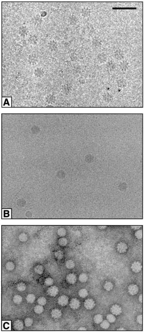
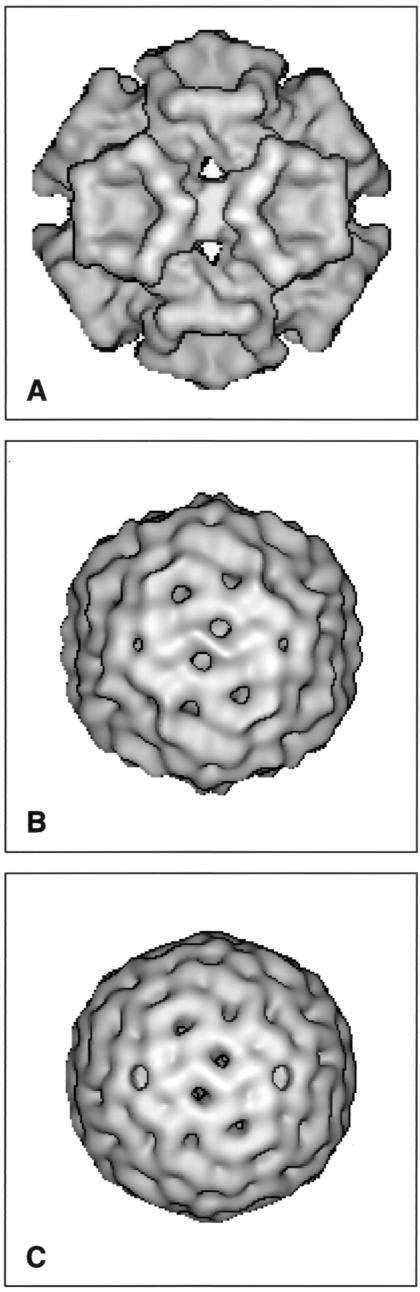
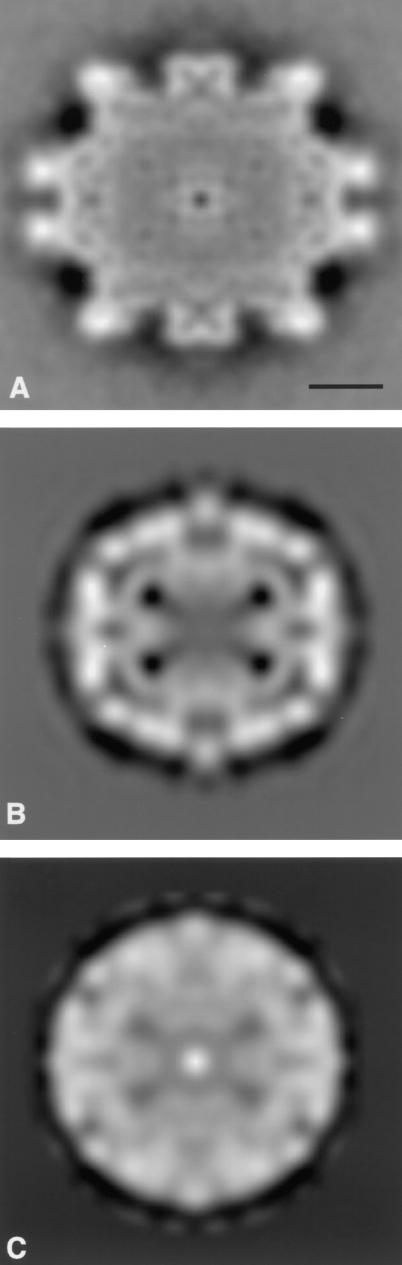
Circoviruses are small, nonenveloped icosahedral animal viruses characterized by circular single-stranded DNA genomes. Their genomes are the smallest possessed by animal viruses. Infections with circoviruses, which can lead to economically important diseases, frequently result in virus-induced damage to lymphoid tissue and immunosuppression. Within the family Circoviridae, different genera are distinguished by differences in genomic organization. Thus, Chicken anemia virus is in the genus Gyrovirus, while porcine circoviruses and Beak and feather disease virus belong to the genus CIRCOVIRUS: Little is known about the structures of circoviruses. Accordingly, we investigated the structures of these three viruses with a view to determining whether they are related. Three-dimensional maps computed from electron micrographs showed that all three viruses have a T=1 organization with capsids formed from 60 subunits. Porcine circovirus type 2 and beak and feather disease virus show similar capsid structures with flat pentameric morphological units, whereas chicken anemia virus has stikingly different protruding pentagonal trumpet-shaped units. It thus appears that the structures of viruses in the same genus are related but that those of viruses in different genera are unrelated.
Figures
References
-
- Allan, G. M., F. McNeilly, S. Kennedy, B. Daft, E. G. Clark, J. A. Ellis, D. M. Haines, B. M. Meehan, and B. M. Adair. 1998. Isolation of porcine circovirus-like virus from pigs with a wasting disease in the United States of America and Europe. J. Vet. Diagn. Investig. 10:3-10. - PubMed
-
- Bassami, M. R., D. Berryman, G. E. Wilcox, and S. R. Raidal. 1998. Psittacine beak and feather disease virus nucleotide sequence analysis and its relationship to porcine circovirus, plant circoviruses, and chicken anemia virus. J. Virol. 249:453-459. - PubMed
-
- Böttcher, B., and R. A. Crowther. 1996. Difference imaging reveals ordered regions of RNA in turnip yellow mosaic virus. Structure 4:387-394. - PubMed
-
- Clark, E. G. 1997. Post weaning multisystemic wasting syndrome, p. 499-501. In Proceedings of the 28th Annual Meeting of the American Association of Swine Practitioners, Quebec City, Quebec, Canada.
-
- Crowther, R. A. 1971. Procedures for three-dimensional reconstruction of spherical viruses by Fourier synthesis from electron micrographs. Philos. Trans. R. Soc. Lond. B 261:221-230. - PubMed
Publication types
MeSH terms
LinkOut - more resources
Full Text Sources
Other Literature Sources
