. 2003 Dec;77(24):13136-45.
doi: 10.1128/jvi.77.24.13136-13145.2003.

Chromosomal integration and homologous gene targeting by replication-incompetent vectors based on the autonomous parvovirus minute virus of mice

Affiliations

Chromosomal integration and homologous gene targeting by replication-incompetent vectors based on the autonomous parvovirus minute virus of mice

Paul C Hendrie et al. J Virol. 2003 Dec.

Abstract

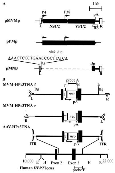
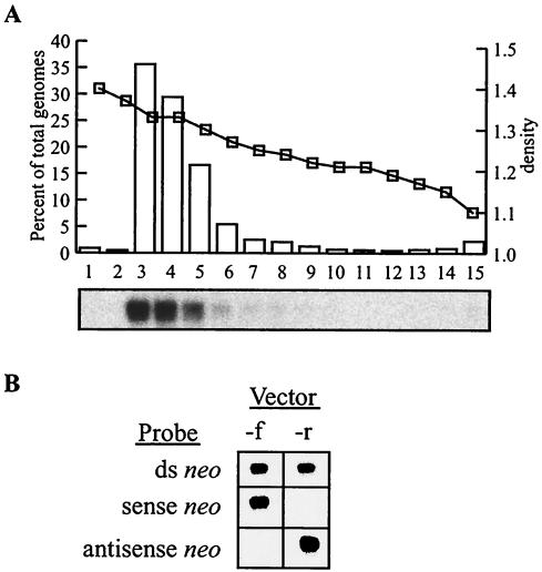
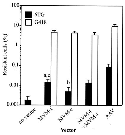
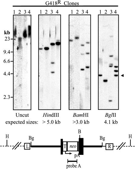
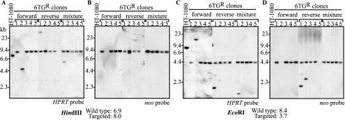
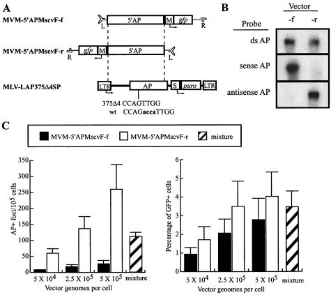
The molecular mechanisms responsible for random integration and gene targeting by recombinant adeno-associated virus (AAV) vectors are largely unknown, and whether vectors derived from autonomous parvoviruses transduce cells by similar pathways has not been investigated. In this report, we constructed vectors based on the autonomous parvovirus minute virus of mice (MVM) that were designed to introduce a neomycin resistance expression cassette (neo) into the X-linked human hypoxanthine phosphoribosyl transferase (HPRT) locus. High-titer, replication-incompetent MVM vector stocks were generated with a two-plasmid transfection system that preserved the wild-type characteristic of packaging only one DNA strand. Vectors with inserts in the forward or reverse orientations packaged noncoding or coding strands, respectively. In human HT-1080 cells, MVM vector random integration frequencies (neo(+) colonies) were comparable to those obtained with AAV vectors, and no difference was observed for noncoding and coding strands. HPRT gene-targeting frequencies (HPRT mutant colonies) were lower with MVM vectors, and the noncoding strand frequency was threefold greater than that of the coding strand. Random integration and gene-targeting events were confirmed by Southern blot analysis of G418- and 6-thioguanine (6TG)-resistant clones. In separate experiments, correction of an alkaline phosphatase (AP) gene by gene targeting was nine times more effective with a coding strand vector. The data suggest that single-stranded parvoviral vector genomes are substrates for gene targeting and possibly for random integration as well.


Figures

FIG. 1.
FIG. 1.
Plasmids and vectors used in this study. (A) Maps of wild-type MVM plasmid pMVMp, packaging plasmid pPMp, and vector backbone plasmid pMNB are shown. The left (L)- and right (R)-terminal palindromes are indicated by open boxes. Solid boxes represent the viral open reading frames for the nonstructural proteins (NS1/2) and capsid proteins (VP1/2). The MVM P4 and P38 promoters are shown with arrows. The location of the first polyadenylation (pA) signal is shown. The location of the cis-acting internal replication sequence (IRS) is shown by an open box below pMVMp. The viral sequence is shown as a thick line. The dashed line represents deleted viral sequence. The sequence of the inserted NS1 nick site is shown above pMNB. (B) Maps of MVM and AAV targeting vectors and the human HPRT locus are shown. Locations the of HPRT exons (black boxes), introns (thick lines), AAV inverted terminal repeats (ITR), MVM left (L)- and right (R)-terminal palindrome sequences, TK promoter (T), neo gene, pA sites, and the neo and HPRT hybridization probes used (probes A and B, respectively) are shown. The AAV and MVM vector terminal repeats are shown in their base-paired secondary structures. Each vector contains the same 4,073-bp targeting cassette composed of 2,915 bp of the HPRT locus sequence with the neo expression cassette disrupting the centrally located exon 3. Relevant endonuclease restriction sites (BamHI, B; BglII, Bg; EcoRI, E; HindIII, H) are shown in panels A and B.
FIG. 2.
FIG. 2.
Characterization of MVM vector stocks. (A) Vector MVM-HPe3TNA-f was purified on an iodixanol gradient, and 2 μl of each fraction was analyzed on an alkaline Southern blot probed for HPRT sequences. The graph depicts the percentage of total genomes present in each fraction (bars) determined by PhosphorImager quantification of the Southern blot (autoradiograph shown below graph), and the calculated density (squares) determined from the RI of each fraction. (B) Strand-specific alkaline Southern blot analysis of MVM-HPe3TNA-f (forward) and MVM-HPe3TNA-r (reverse) vector stocks (3 × 10⁸ genomes each) hybridized with a double-stranded neo probe (top row), a T7 sense neo transcript (middle row), or a T3 antisense neo transcript (bottom row).
FIG. 3.
FIG. 3.
Transduction frequencies of MVM and AAV vectors. A total of 4 × 10⁴ HT-1080 cells were transduced with the indicated vector preparation (2 × 10¹⁰ particles of MVM-HPe3TNA-f [MVM-f] or MVM-HPe3TNA-r [MVM-r], 1 × 10¹⁰ particles of both MVM-f and MVM-r, or 2 × 10⁹ particles of AAV-HPe3TNA [AAV]) and expanded for 10 days, and then the percentage of G418- or 6TG-resistant cells was determined by plating portions in G418, 6TG, or nonselective medium (see Materials and Methods). The G418 (open bars) and 6TG (solid bars) resistance frequencies were determined by dividing the number of G418- or 6TG-resistant colonies obtained by the number of unselected colonies obtained. The mean and standard deviation of five experiments (MVM vector infections) or four experiments (AAV infections) are shown. The P values for student t tests comparing 6TG resistance frequencies of MVM-f to those of the no-vector control (a), MVM-r versus control (b), and MVM-f versus MVM-r (c) were P < 0.005, P = 0.069, and P = 0.015, respectively.
FIG. 4.
FIG. 4.
Southern blot analysis of G418-resistant HT-1080 clones. Genomic DNAs from four representative G418-resistant clones were isolated on 1% agarose gels without digestion or after digestion with HindIII, BamHI or BglII, and probed for neo sequences. The positions of size standards and the expected 4.1-kb BglII vector fragment are shown to the left and right of the panels, respectively. The predicted structure of an integrated provirus is shown below the panels with the locations of HPRT intron sequences (thick line), HPRT exon 3 (dark boxes), MVM right (R)- and left (L)-terminal palindromes, TK promoter (T), neo gene, pA sites, and relevant restriction sites (BamHI, B; BglII, Bg; HindIII, H) indicated.
FIG. 5.
FIG. 5.
Southern blot analysis of 6TG-resistant HT-1080 clones. Genomic DNAs from 15 6TG-resistant clones (5 each from cells transduced with MVM-HPe3TNA-f, MVM-HPe3TNA-r, and the mixture of vectors) and untransduced, parental HT-1080 cells were digested with HindIII (A and B) or EcoRI (C and D) and probed for HPRT (A and C) or neo (B and D) sequences. The positions of size standards are shown to the left of each panel, and the predicted fragment sizes of wild-type and targeted alleles are indicated. The locations of restriction sites and the probe fragments are shown in Fig. 1.
FIG. 6.
FIG. 6.
Replication center assay. NB324K cells were left uninfected (control) or were infected with the indicated amounts of genome-containing particles of wild-type MVM and 108 MVM vector genomes, cultured for 2 days, aspirated onto nylon membrane filters, lysed, and then hybridized to a 5′ NS1 probe. Radioactive spots represent individual cells with replicated NS1-containing genomes due to infection with RCV.
FIG. 7.
FIG. 7.
Determination of the intracellular form of MVM vector genomes. Shown are the results of Southern blot analysis of Hirt supernatants from 4 × 10⁴ HT-1080 cells infected with 2 × 10⁹ genome-containing particles of MVM-HPe3TNA-f prepared 0.5, 24, and 72 h after infection. One-fourth of each sample was run on 0.8% alkaline agarose gels, transferred to nylon membranes, and hybridized with a double-stranded neo probe (A) or a single-stranded antisense neo probe (B). Arrows to the right of each panel show expected locations of single-stranded monomer (m) and dimer (d) genomes.
FIG. 8.
FIG. 8.
AP gene-targeting experiments. (A) Diagrams of MVM and retroviral vectors for AP gene targeting are shown. The murine leukemia virus (MLV) retroviral vector LAP375Δ4SP is shown with the positions of the LTR, AP gene, SV40 promoter (S), and puromycin resistance gene (puro) indicated. The 4-bp deletion in AP is shown below the AP gene along with the corresponding wild-type (wt) sequence: the deleted bases are lowercase and boldface. Maps of MVM targeting vectors containing 5′ portions of the AP gene are shown above the MLV-LAP375Δ4SP target site, with the locations of left (L)- and right (R)-terminal palindrome sequences, murine stem cell virus LTR promoter (M), and green fluorescent protein (gfp) indicated. Each targeting vector contains 2,498 bp of homology to the target sequence, as indicated by dashed lines. (B) Strand-specific alkaline Southern blot analysis of MVM-5′APMscvF-f (forward) and MVM-5′APMscvF-r (reverse) vector stocks (1 × 10⁹ genomes each) hybridized with a double-stranded AP probe (top row), a T7 sense AP transcript (middle row), or a T3 antisense AP transcript (bottom row). (C) AP gene-targeting rates and GFP gene addition rates are shown for MVM-5′APMscvF-f (solid bars), MVM-5′APMscvF-r (open bars), and a mixture of both vectors (hatched bars) in HT/LAP375Δ4SPc9 cells at the indicated MOIs. The mixture contained 2.5 × 10⁵ genomes of forward and reverse vector stocks. Mean values are shown with standard deviations from three independent experiments.

Similar articles

Cited by

References

    1. Aitken, M. L., R. B. Moss, D. A. Waltz, M. E. Dovey, M. R. Tonelli, S. C. McNamara, R. L. Gibson, B. W. Ramsey, B. J. Carter, and T. C. Reynolds. 2001. A phase I study of aerosolized administration of tgAAVCF to cystic fibrosis subjects with mild lung disease. Hum. Gene Ther. 12:1907-1916. - PubMed
    1. Avalosse, B. L., F. Dupont, P. Spegelaere, N. Mine, and A. Burny. 1996. Method for concentrating and purifying recombinant autonomous parvovirus vectors designed for tumour-cell-targeted gene therapy. J. Virol. Methods 62:179-183. - PubMed
    1. Berns, K. I. 1996. Parvoviridae: the viruses and their replication, p. 2173-2197. In B. N. Fields, D. M. Knipe, P. M. Howley et al. (ed.), Fields virology, 3rd ed. Lippincott-Raven Publishers, Philadelphia, Pa.
    1. Berns, K. I., and S. Adler. 1972. Separation of two types of adeno-associated virus particles containing complementary polynucleotide chains. J. Virol. 9:394-396. - PMC - PubMed
    1. Bourguignon, G. J., P. J. Tattersall, and D. C. Ward. 1976. DNA of minute virus of mice: self-priming, nonpermuted, single-stranded genome with a 5′-terminal hairpin duplex. J. Virol. 20:290-306. - PMC - PubMed

Publication types

Substances

LinkOut - more resources