

Lytic viral replication as a contributor to the detection of Epstein-Barr virus in breast cancer
- PMID: 14645583
- PMCID: PMC296054
- DOI: 10.1128/jvi.77.24.13267-13274.2003
Lytic viral replication as a contributor to the detection of Epstein-Barr virus in breast cancer
Abstract
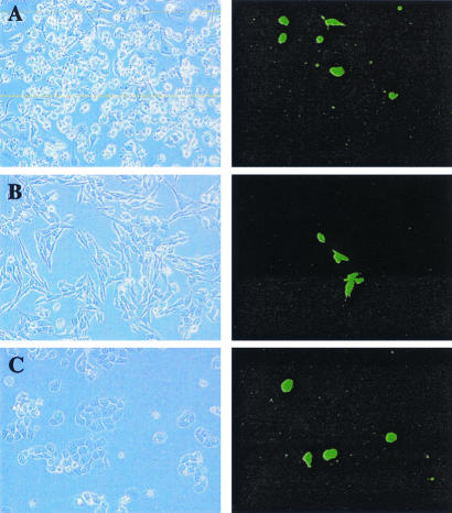
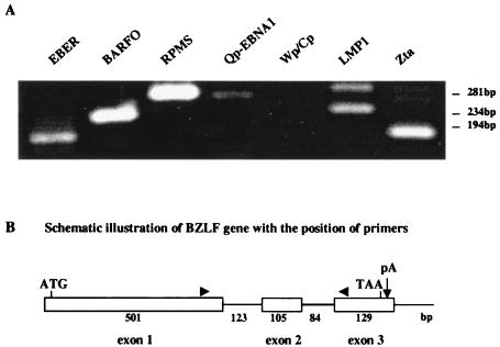
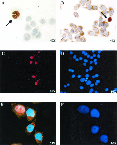
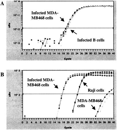
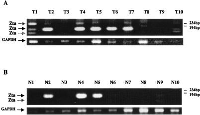
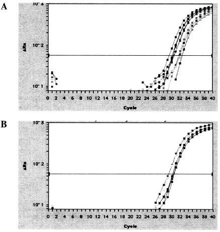
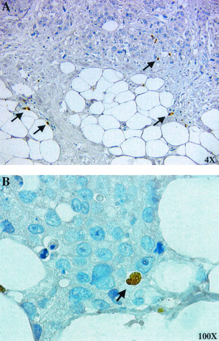
Epstein-Barr virus (EBV) has an accepted association with the epithelial malignancy nasopharyngeal carcinoma and has also been reported in other more controversial carcinoma settings. Evaluation of EBV association with epithelial carcinomas such as breast cancer would benefit from a better understanding of the outcome of EBV infection of these cells. Cell-free preparations of a green fluorescent protein-expressing virus, BX1, were used to infect breast cancer cell lines, which were then examined for EBV gene expression and viral genome copy number. Reverse transcription-PCR analyses revealed that the cells supported a mixture of latency II and lytic EBV gene expression. Lytic Zta and BMRF1 protein expression was detected by immunohistochemistry, and DNA PCR analyses estimated an EBV copy number of 300 to 600 genomes per infected cell. Evidence for lytic EBV expression was also found in breast tissue, where reverse transcription-PCR analyses detected lytic Zta transcripts in 7 of 10 breast carcinoma tissues and 4 of 10 normal tissues from the same patients. Scattered cells immunoreactive for Zta protein were also detectable in breast carcinoma. Quantitative real-time PCR analysis of EBV-positive breast carcinoma tissues suggested that less than 0.1% of the cells contained viral genomes. We suggest that sporadic lytic EBV infection may contribute to PCR-based detection of EBV in traditionally nonvirally associated epithelial malignancies.
Figures
Similar articles
-
The SWI/SNF Chromatin Regulator BRG1 Modulates the Transcriptional Regulatory Activity of the Epstein-Barr Virus DNA Polymerase Processivity Factor BMRF1.J Virol. 2017 Apr 13;91(9):e02114-16. doi: 10.1128/JVI.02114-16. Print 2017 May 1. J Virol. 2017. PMID: 28228591 Free PMC article.
-
Contribution of C/EBP proteins to Epstein-Barr virus lytic gene expression and replication in epithelial cells.J Virol. 2006 Feb;80(3):1098-109. doi: 10.1128/JVI.80.3.1098-1109.2006. J Virol. 2006. PMID: 16414987 Free PMC article.
-
Efficient Translation of Epstein-Barr Virus (EBV) DNA Polymerase Contributes to the Enhanced Lytic Replication Phenotype of M81 EBV.J Virol. 2018 Feb 26;92(6):e01794-17. doi: 10.1128/JVI.01794-17. Print 2018 Mar 15. J Virol. 2018. PMID: 29263273 Free PMC article.
-
Latent and lytic Epstein-Barr virus replication strategies.Rev Med Virol. 2005 Jan-Feb;15(1):3-15. doi: 10.1002/rmv.441. Rev Med Virol. 2005. PMID: 15386591 Review.
-
Regulation of cellular and viral protein expression by the Epstein-Barr virus transcriptional regulator Zta: implications for therapy of EBV associated tumors.Cancer Biol Ther. 2009 Jun;8(11):987-95. doi: 10.4161/cbt.8.11.8369. Epub 2009 Jun 10. Cancer Biol Ther. 2009. PMID: 19448399 Review.
Cited by
-
CRISPR/Cas13-Mediated Inhibition of EBNA1 for Suppression of Epstein-Barr Virus Transcripts and DNA Load in Nasopharyngeal Carcinoma Cells.Viruses. 2025 Jun 26;17(7):899. doi: 10.3390/v17070899. Viruses. 2025. PMID: 40733516 Free PMC article.
-
Epstein-Barr virus infection in ex vivo tonsil epithelial cell cultures of asymptomatic carriers.J Virol. 2004 Nov;78(22):12613-24. doi: 10.1128/JVI.78.22.12613-12624.2004. J Virol. 2004. PMID: 15507648 Free PMC article.
-
Epstein-Barr virus as a marker of biological aggressiveness in breast cancer.Br J Cancer. 2011 Jan 18;104(2):332-7. doi: 10.1038/sj.bjc.6606048. Epub 2010 Dec 21. Br J Cancer. 2011. PMID: 21179039 Free PMC article.
-
Dysregulation of HER2/HER3 signaling axis in Epstein-Barr virus-infected breast carcinoma cells.J Virol. 2007 Jun;81(11):5705-13. doi: 10.1128/JVI.00076-07. Epub 2007 Mar 21. J Virol. 2007. PMID: 17376931 Free PMC article.
-
Oncogenic Properties of the EBV ZEBRA Protein.Cancers (Basel). 2020 Jun 5;12(6):1479. doi: 10.3390/cancers12061479. Cancers (Basel). 2020. PMID: 32517128 Free PMC article. Review.
References
-
- Babcock, G. L., L. L. Decker, M. Volk, and D. A. Thorley-Lawson. 1998. EBV persistence in memory B cells in vivo. Immunity 9:395-404. - PubMed
-
- Barel, M., M. Le Romancer, and R. Frade. 2001. Activation of the EBV/C3d receptor (CR2, CD21) on human B lymphocyte surface triggers tyrosine phosphorylation of the 95-kDa nucleolin and its interaction with phosphatidylinositol 3 kinase. J. Immunol. 166:3167-3173. - PubMed
-
- Bonnet, M., J. M. Guinebretiere, E. Kremmer, V. Grunewald, E. Benhamou, G. Contesso, and I. Joab. 1999. Detection of Epstein-Barr virus in invasive breast cancers. J. Natl. Cancer Inst. 91:1376-1381. - PubMed
-
- Bowman, T., R. Garcia, J. Turkson, and R. Jove. 2000. STATs in oncogenesis. Oncogene 19:2474-2488. - PubMed
Publication types
MeSH terms
Substances
Grants and funding
LinkOut - more resources
Full Text Sources
Medical
